Manganese »
PDB 2feu-2hr6 »
2hk1 »
Manganese in PDB 2hk1: Crystal Structure of D-Psicose 3-Epimerase (Dpease) in the Presence of D-Fructose
Protein crystallography data
The structure of Crystal Structure of D-Psicose 3-Epimerase (Dpease) in the Presence of D-Fructose, PDB code: 2hk1
was solved by
K.Kim,
H.J.Kim,
D.K.Oh,
S.S.Cha,
S.Rhee,
with X-Ray Crystallography technique. A brief refinement statistics is given in the table below:
| Resolution Low / High (Å) | 33.50 / 2.30 |
| Space group | P 21 21 21 |
| Cell size a, b, c (Å), α, β, γ (°) | 103.900, 113.500, 133.800, 90.00, 90.00, 90.00 |
| R / Rfree (%) | 19.2 / 23.9 |
Manganese Binding Sites:
The binding sites of Manganese atom in the Crystal Structure of D-Psicose 3-Epimerase (Dpease) in the Presence of D-Fructose
(pdb code 2hk1). This binding sites where shown within
5.0 Angstroms radius around Manganese atom.
In total 4 binding sites of Manganese where determined in the Crystal Structure of D-Psicose 3-Epimerase (Dpease) in the Presence of D-Fructose, PDB code: 2hk1:
Jump to Manganese binding site number: 1; 2; 3; 4;
In total 4 binding sites of Manganese where determined in the Crystal Structure of D-Psicose 3-Epimerase (Dpease) in the Presence of D-Fructose, PDB code: 2hk1:
Jump to Manganese binding site number: 1; 2; 3; 4;
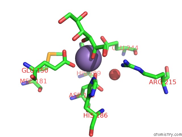
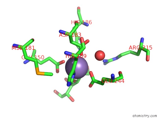
Manganese binding site 1 out of 4 in 2hk1
Go back to
Manganese binding site 1 out
of 4 in the Crystal Structure of D-Psicose 3-Epimerase (Dpease) in the Presence of D-Fructose
Mono view
Stereo pair view
Mono view
Stereo pair view
A full contact list of Manganese with other atoms in the Mn binding
site number 1 of Crystal Structure of D-Psicose 3-Epimerase (Dpease) in the Presence of D-Fructose within 5.0Å range:
|
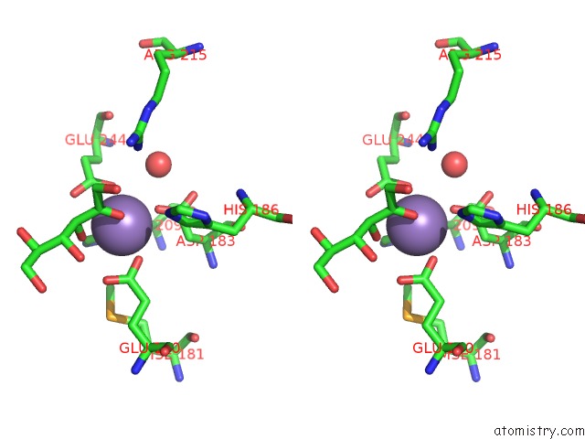
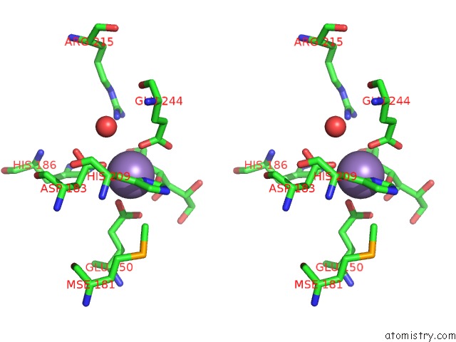
Manganese binding site 2 out of 4 in 2hk1
Go back to
Manganese binding site 2 out
of 4 in the Crystal Structure of D-Psicose 3-Epimerase (Dpease) in the Presence of D-Fructose
Mono view
Stereo pair view
Mono view
Stereo pair view
A full contact list of Manganese with other atoms in the Mn binding
site number 2 of Crystal Structure of D-Psicose 3-Epimerase (Dpease) in the Presence of D-Fructose within 5.0Å range:
|
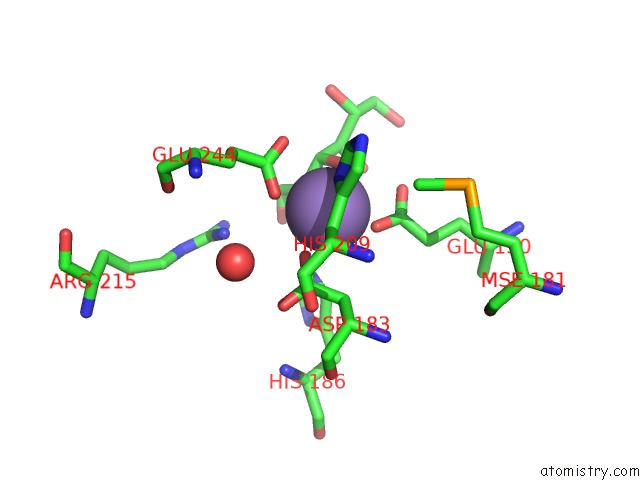
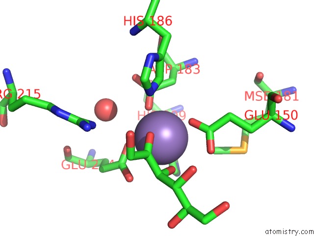
Manganese binding site 3 out of 4 in 2hk1
Go back to
Manganese binding site 3 out
of 4 in the Crystal Structure of D-Psicose 3-Epimerase (Dpease) in the Presence of D-Fructose
Mono view
Stereo pair view
Mono view
Stereo pair view
A full contact list of Manganese with other atoms in the Mn binding
site number 3 of Crystal Structure of D-Psicose 3-Epimerase (Dpease) in the Presence of D-Fructose within 5.0Å range:
|
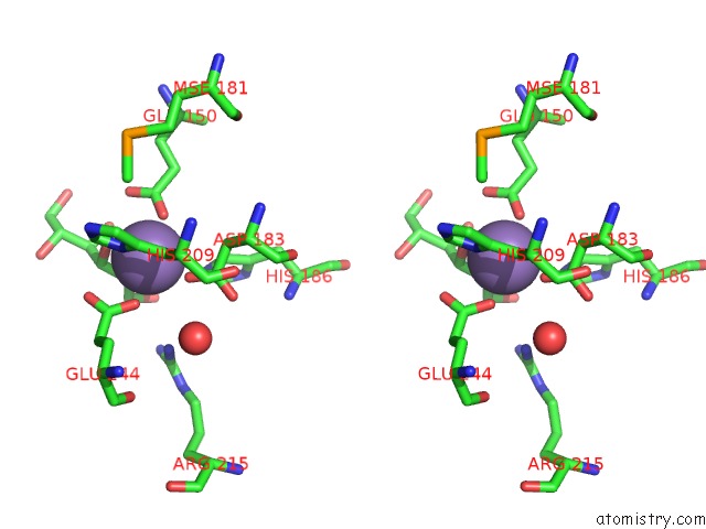
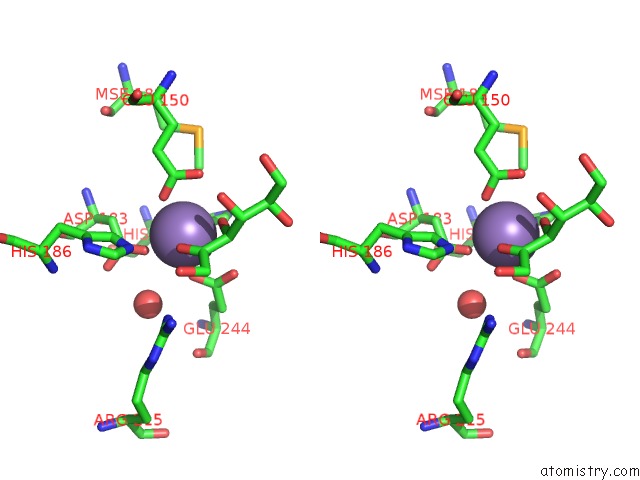
Manganese binding site 4 out of 4 in 2hk1
Go back to
Manganese binding site 4 out
of 4 in the Crystal Structure of D-Psicose 3-Epimerase (Dpease) in the Presence of D-Fructose
Mono view
Stereo pair view
Mono view
Stereo pair view
A full contact list of Manganese with other atoms in the Mn binding
site number 4 of Crystal Structure of D-Psicose 3-Epimerase (Dpease) in the Presence of D-Fructose within 5.0Å range:
|
Reference:
K.Kim,
H.J.Kim,
D.K.Oh,
S.S.Cha,
S.Rhee.
Crystal Structure of D-Psicose 3-Epimerase From Agrobacterium Tumefaciens and Its Complex with True Substrate D-Fructose: A Pivotal Role of Metal in Catalysis, An Active Site For the Non-Phosphorylated Substrate, and Its Conformational Changes J.Mol.Biol. V. 361 920 2006.
ISSN: ISSN 0022-2836
PubMed: 16876192
DOI: 10.1016/J.JMB.2006.06.069
Page generated: Sat Oct 5 14:17:52 2024
ISSN: ISSN 0022-2836
PubMed: 16876192
DOI: 10.1016/J.JMB.2006.06.069
Last articles
Fe in 7ZXYFe in 7ZZL
Fe in 7ZXX
Fe in 7ZXL
Fe in 7ZXJ
Fe in 7ZXC
Fe in 7ZXD
Fe in 7ZX5
Fe in 7ZX6
Fe in 7ZX3