Manganese »
PDB 2feu-2hr6 »
2gdf »
Manganese in PDB 2gdf: Crystal Structure of Dioclea Violacea Seed Lectin
Protein crystallography data
The structure of Crystal Structure of Dioclea Violacea Seed Lectin, PDB code: 2gdf
was solved by
F.Gallego Del Sol,
J.J.C.Chornet,
B.S.Cavada,
with X-Ray Crystallography technique. A brief refinement statistics is given in the table below:
| Resolution Low / High (Å) | 45.00 / 2.40 |
| Space group | P 32 |
| Cell size a, b, c (Å), α, β, γ (°) | 72.938, 72.938, 161.362, 90.00, 90.00, 120.00 |
| R / Rfree (%) | 22.7 / 25.8 |
Other elements in 2gdf:
The structure of Crystal Structure of Dioclea Violacea Seed Lectin also contains other interesting chemical elements:
| Calcium | (Ca) | 2 atoms |
Manganese Binding Sites:
The binding sites of Manganese atom in the Crystal Structure of Dioclea Violacea Seed Lectin
(pdb code 2gdf). This binding sites where shown within
5.0 Angstroms radius around Manganese atom.
In total 2 binding sites of Manganese where determined in the Crystal Structure of Dioclea Violacea Seed Lectin, PDB code: 2gdf:
Jump to Manganese binding site number: 1; 2;
In total 2 binding sites of Manganese where determined in the Crystal Structure of Dioclea Violacea Seed Lectin, PDB code: 2gdf:
Jump to Manganese binding site number: 1; 2;
Manganese binding site 1 out of 2 in 2gdf
Go back to
Manganese binding site 1 out
of 2 in the Crystal Structure of Dioclea Violacea Seed Lectin
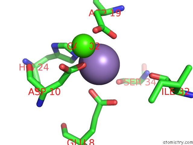
Mono view
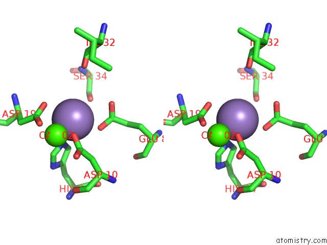
Stereo pair view
Mono view
Stereo pair view
A full contact list of Manganese with other atoms in the Mn binding
site number 1 of Crystal Structure of Dioclea Violacea Seed Lectin within 5.0Å range:
|
Manganese binding site 2 out of 2 in 2gdf
Go back to
Manganese binding site 2 out
of 2 in the Crystal Structure of Dioclea Violacea Seed Lectin
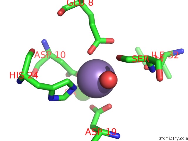
Mono view
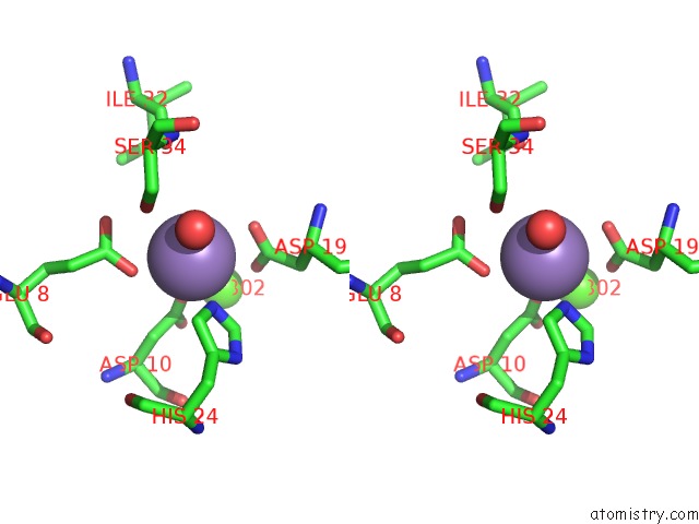
Stereo pair view
Mono view
Stereo pair view
A full contact list of Manganese with other atoms in the Mn binding
site number 2 of Crystal Structure of Dioclea Violacea Seed Lectin within 5.0Å range:
|
Reference:
F.Gallego Del Sol,
J.J.C.Chornet,
B.S.Cavada.
Crystal Structure of Dioclea Violacea Seed Lectin To Be Published.
Page generated: Sat Oct 5 14:10:05 2024
Last articles
Fe in 8AA7Fe in 8A9P
Fe in 8A91
Fe in 8A90
Fe in 8A8L
Fe in 8A82
Fe in 8A5G
Fe in 8A6W
Fe in 8A5F
Fe in 8A1Y