Manganese »
PDB 2dvd-2fer »
2e7t »
Manganese in PDB 2e7t: Crystal Structure of Basic Winged Bean Lectin in Complex with A Blood Group Trisaccharide
Protein crystallography data
The structure of Crystal Structure of Basic Winged Bean Lectin in Complex with A Blood Group Trisaccharide, PDB code: 2e7t
was solved by
K.A.Kulkarni,
S.Katiyar,
A.Surolia,
M.Vijayan,
K.Suguna,
with X-Ray Crystallography technique. A brief refinement statistics is given in the table below:
| Resolution Low / High (Å) | 29.00 / 2.65 |
| Space group | P 21 21 2 |
| Cell size a, b, c (Å), α, β, γ (°) | 157.729, 91.130, 73.718, 90.00, 90.00, 90.00 |
| R / Rfree (%) | 19.2 / 24.4 |
Other elements in 2e7t:
The structure of Crystal Structure of Basic Winged Bean Lectin in Complex with A Blood Group Trisaccharide also contains other interesting chemical elements:
| Calcium | (Ca) | 4 atoms |
Manganese Binding Sites:
The binding sites of Manganese atom in the Crystal Structure of Basic Winged Bean Lectin in Complex with A Blood Group Trisaccharide
(pdb code 2e7t). This binding sites where shown within
5.0 Angstroms radius around Manganese atom.
In total 4 binding sites of Manganese where determined in the Crystal Structure of Basic Winged Bean Lectin in Complex with A Blood Group Trisaccharide, PDB code: 2e7t:
Jump to Manganese binding site number: 1; 2; 3; 4;
In total 4 binding sites of Manganese where determined in the Crystal Structure of Basic Winged Bean Lectin in Complex with A Blood Group Trisaccharide, PDB code: 2e7t:
Jump to Manganese binding site number: 1; 2; 3; 4;
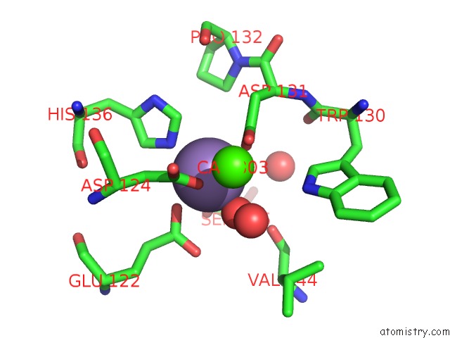
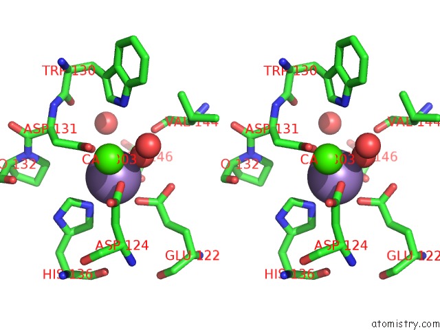
Manganese binding site 1 out of 4 in 2e7t
Go back to
Manganese binding site 1 out
of 4 in the Crystal Structure of Basic Winged Bean Lectin in Complex with A Blood Group Trisaccharide
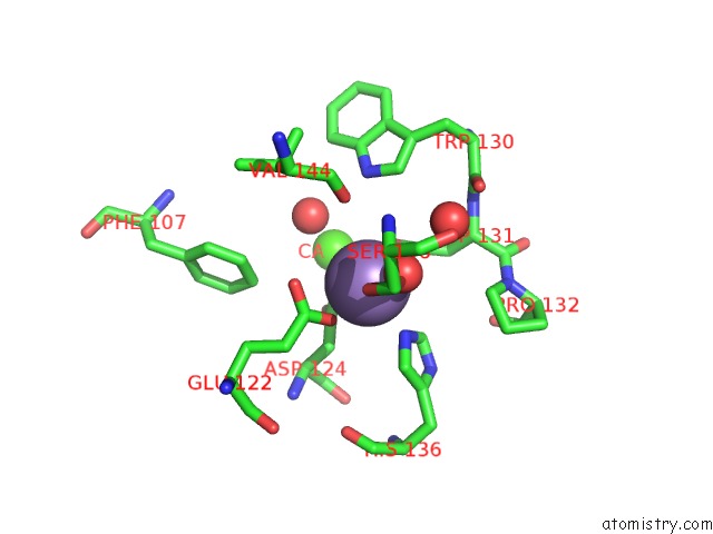
Mono view
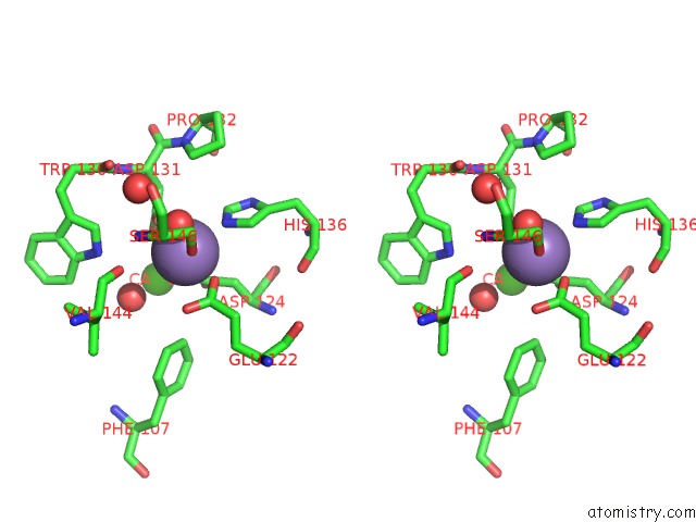
Stereo pair view
Mono view
Stereo pair view
A full contact list of Manganese with other atoms in the Mn binding
site number 1 of Crystal Structure of Basic Winged Bean Lectin in Complex with A Blood Group Trisaccharide within 5.0Å range:
|
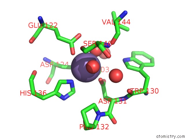
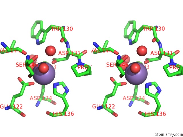
Manganese binding site 2 out of 4 in 2e7t
Go back to
Manganese binding site 2 out
of 4 in the Crystal Structure of Basic Winged Bean Lectin in Complex with A Blood Group Trisaccharide
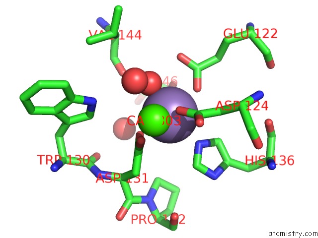
Mono view
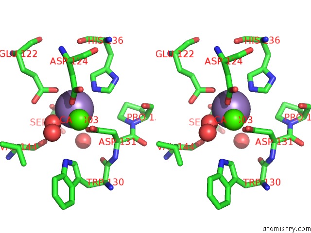
Stereo pair view
Mono view
Stereo pair view
A full contact list of Manganese with other atoms in the Mn binding
site number 2 of Crystal Structure of Basic Winged Bean Lectin in Complex with A Blood Group Trisaccharide within 5.0Å range:
|
Manganese binding site 3 out of 4 in 2e7t
Go back to
Manganese binding site 3 out
of 4 in the Crystal Structure of Basic Winged Bean Lectin in Complex with A Blood Group Trisaccharide
Mono view
Stereo pair view
Mono view
Stereo pair view
A full contact list of Manganese with other atoms in the Mn binding
site number 3 of Crystal Structure of Basic Winged Bean Lectin in Complex with A Blood Group Trisaccharide within 5.0Å range:
|
Manganese binding site 4 out of 4 in 2e7t
Go back to
Manganese binding site 4 out
of 4 in the Crystal Structure of Basic Winged Bean Lectin in Complex with A Blood Group Trisaccharide
Mono view
Stereo pair view
Mono view
Stereo pair view
A full contact list of Manganese with other atoms in the Mn binding
site number 4 of Crystal Structure of Basic Winged Bean Lectin in Complex with A Blood Group Trisaccharide within 5.0Å range:
|
Reference:
K.A.Kulkarni,
S.Katiyar,
A.Surolia,
M.Vijayan,
K.Suguna.
Generation of Blood Group Specificity: New Insights From Structural Studies on the Complexes of A- and B-Reactive Saccharides with Basic Winged Bean Agglutinin Proteins V. 68 762 2007.
ISSN: ISSN 0887-3585
PubMed: 17510954
DOI: 10.1002/PROT.21428
Page generated: Sat Oct 5 13:57:40 2024
ISSN: ISSN 0887-3585
PubMed: 17510954
DOI: 10.1002/PROT.21428
Last articles
I in 5R4DI in 5S40
I in 5R4B
I in 5R4C
I in 5R4A
I in 5R49
I in 5OSW
I in 5R47
I in 5R48
I in 5R46