Manganese »
PDB 2cev-2dvb »
2dti »
Manganese in PDB 2dti: Crystal Structure of Biotin Protein Ligase From Pyrococcus Horikoshii OT3 in Complex with Biotinyl-5'-Amp, Pyrophosphate and Mn(2+)
Enzymatic activity of Crystal Structure of Biotin Protein Ligase From Pyrococcus Horikoshii OT3 in Complex with Biotinyl-5'-Amp, Pyrophosphate and Mn(2+)
All present enzymatic activity of Crystal Structure of Biotin Protein Ligase From Pyrococcus Horikoshii OT3 in Complex with Biotinyl-5'-Amp, Pyrophosphate and Mn(2+):
6.3.4.15;
6.3.4.15;
Protein crystallography data
The structure of Crystal Structure of Biotin Protein Ligase From Pyrococcus Horikoshii OT3 in Complex with Biotinyl-5'-Amp, Pyrophosphate and Mn(2+), PDB code: 2dti
was solved by
B.Bagautdinov,
N.Kunishima,
Riken Structural Genomics/Proteomicsinitiative (Rsgi),
with X-Ray Crystallography technique. A brief refinement statistics is given in the table below:
| Resolution Low / High (Å) | 35.54 / 2.20 |
| Space group | P 1 21 1 |
| Cell size a, b, c (Å), α, β, γ (°) | 38.351, 82.628, 72.718, 90.00, 102.22, 90.00 |
| R / Rfree (%) | 23.4 / 28.4 |
Manganese Binding Sites:
The binding sites of Manganese atom in the Crystal Structure of Biotin Protein Ligase From Pyrococcus Horikoshii OT3 in Complex with Biotinyl-5'-Amp, Pyrophosphate and Mn(2+)
(pdb code 2dti). This binding sites where shown within
5.0 Angstroms radius around Manganese atom.
In total 2 binding sites of Manganese where determined in the Crystal Structure of Biotin Protein Ligase From Pyrococcus Horikoshii OT3 in Complex with Biotinyl-5'-Amp, Pyrophosphate and Mn(2+), PDB code: 2dti:
Jump to Manganese binding site number: 1; 2;
In total 2 binding sites of Manganese where determined in the Crystal Structure of Biotin Protein Ligase From Pyrococcus Horikoshii OT3 in Complex with Biotinyl-5'-Amp, Pyrophosphate and Mn(2+), PDB code: 2dti:
Jump to Manganese binding site number: 1; 2;
Manganese binding site 1 out of 2 in 2dti
Go back to
Manganese binding site 1 out
of 2 in the Crystal Structure of Biotin Protein Ligase From Pyrococcus Horikoshii OT3 in Complex with Biotinyl-5'-Amp, Pyrophosphate and Mn(2+)
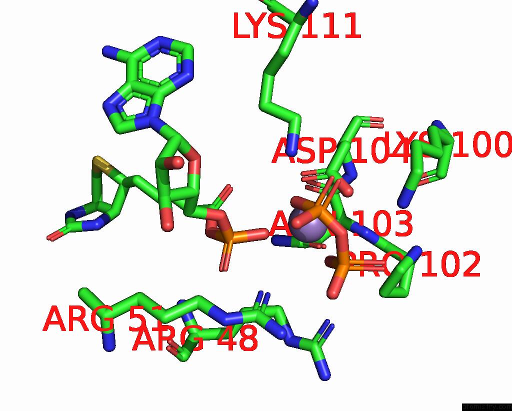
Mono view
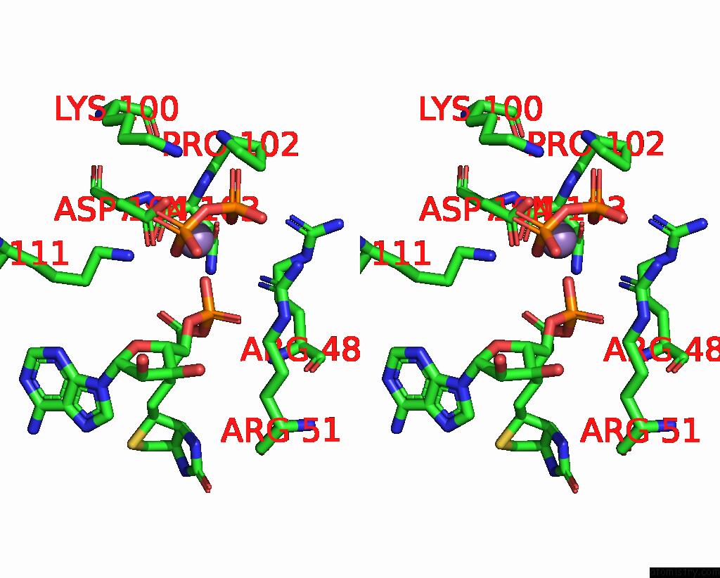
Stereo pair view
Mono view
Stereo pair view
A full contact list of Manganese with other atoms in the Mn binding
site number 1 of Crystal Structure of Biotin Protein Ligase From Pyrococcus Horikoshii OT3 in Complex with Biotinyl-5'-Amp, Pyrophosphate and Mn(2+) within 5.0Å range:
|
Manganese binding site 2 out of 2 in 2dti
Go back to
Manganese binding site 2 out
of 2 in the Crystal Structure of Biotin Protein Ligase From Pyrococcus Horikoshii OT3 in Complex with Biotinyl-5'-Amp, Pyrophosphate and Mn(2+)
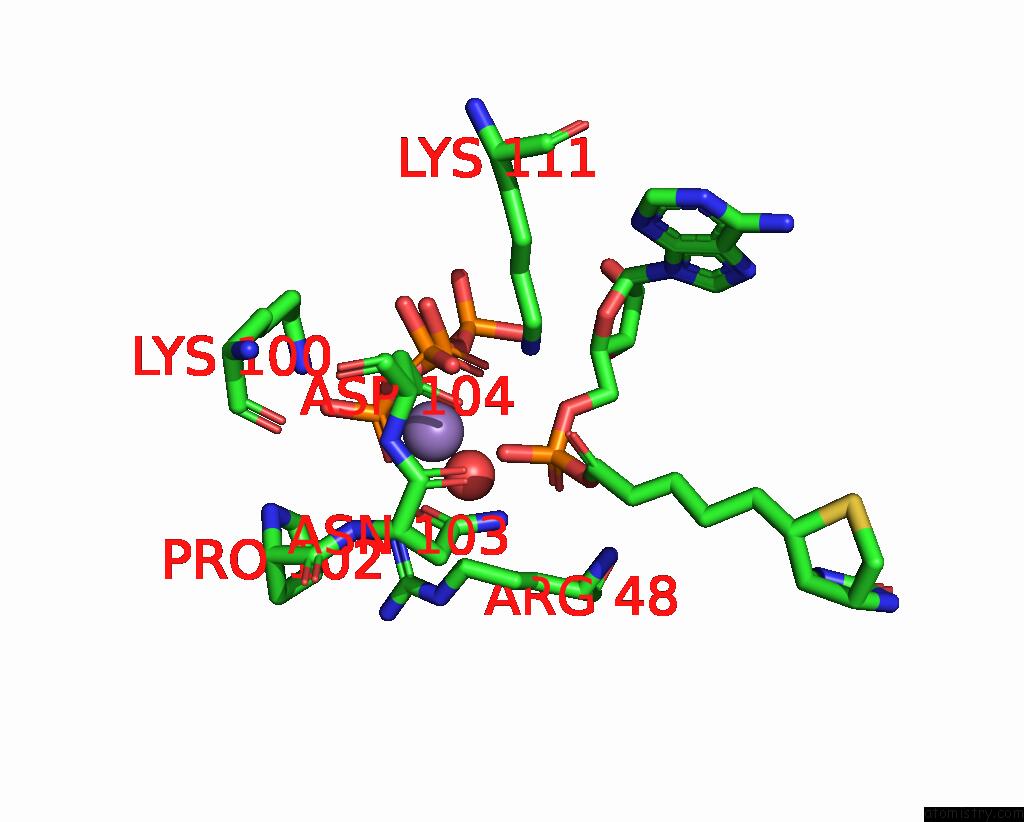
Mono view
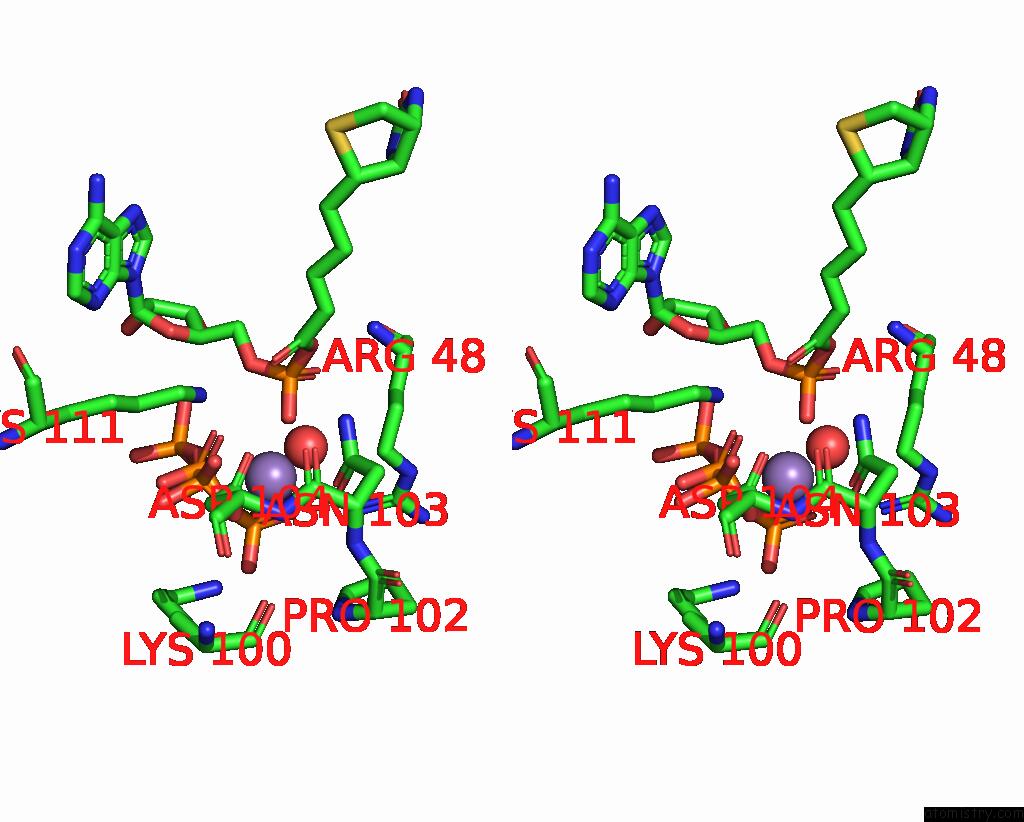
Stereo pair view
Mono view
Stereo pair view
A full contact list of Manganese with other atoms in the Mn binding
site number 2 of Crystal Structure of Biotin Protein Ligase From Pyrococcus Horikoshii OT3 in Complex with Biotinyl-5'-Amp, Pyrophosphate and Mn(2+) within 5.0Å range:
|
Reference:
B.Bagautdinov,
N.Kunishima.
Ligand Structures of Biotin Protein Ligase From Pyrococcus Horikoshii OT3 To Be Published.
Page generated: Sat Oct 5 13:46:49 2024
Last articles
I in 8KG4I in 8KB2
I in 8KEU
I in 8K4Z
I in 8K0T
I in 8JLN
I in 8J9S
I in 8JLJ
I in 8IKP
I in 8IKS