Manganese »
PDB 2ayl-2ce4 »
2bcd »
Manganese in PDB 2bcd: X-Ray Crystal Structure of Protein Phosphatase-1 with the Marine Toxin Motuporin Bound
Enzymatic activity of X-Ray Crystal Structure of Protein Phosphatase-1 with the Marine Toxin Motuporin Bound
All present enzymatic activity of X-Ray Crystal Structure of Protein Phosphatase-1 with the Marine Toxin Motuporin Bound:
3.1.3.16;
3.1.3.16;
Protein crystallography data
The structure of X-Ray Crystal Structure of Protein Phosphatase-1 with the Marine Toxin Motuporin Bound, PDB code: 2bcd
was solved by
J.T.Maynes,
H.A.Luu,
M.M.Cherney,
R.J.Andersen,
D.Williams,
C.F.Holmes,
M.N.James,
with X-Ray Crystallography technique. A brief refinement statistics is given in the table below:
| Resolution Low / High (Å) | 31.75 / 2.10 |
| Space group | P 42 21 2 |
| Cell size a, b, c (Å), α, β, γ (°) | 100.955, 100.955, 63.485, 90.00, 90.00, 90.00 |
| R / Rfree (%) | 22.1 / 26.4 |
Manganese Binding Sites:
The binding sites of Manganese atom in the X-Ray Crystal Structure of Protein Phosphatase-1 with the Marine Toxin Motuporin Bound
(pdb code 2bcd). This binding sites where shown within
5.0 Angstroms radius around Manganese atom.
In total 8 binding sites of Manganese where determined in the X-Ray Crystal Structure of Protein Phosphatase-1 with the Marine Toxin Motuporin Bound, PDB code: 2bcd:
Jump to Manganese binding site number: 1; 2; 3; 4; 5; 6; 7; 8;
In total 8 binding sites of Manganese where determined in the X-Ray Crystal Structure of Protein Phosphatase-1 with the Marine Toxin Motuporin Bound, PDB code: 2bcd:
Jump to Manganese binding site number: 1; 2; 3; 4; 5; 6; 7; 8;
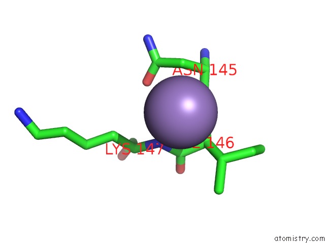
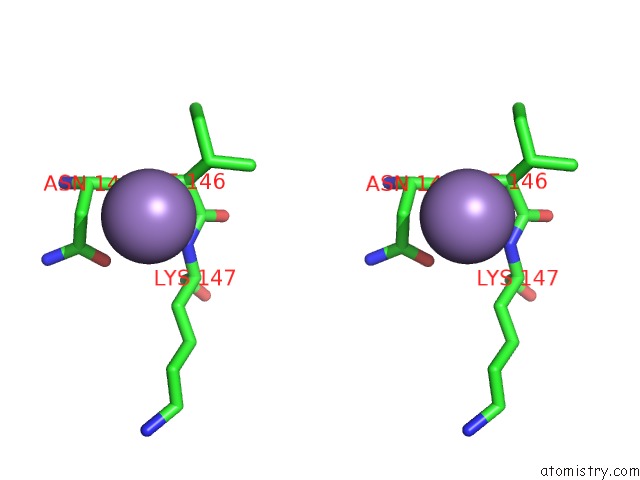
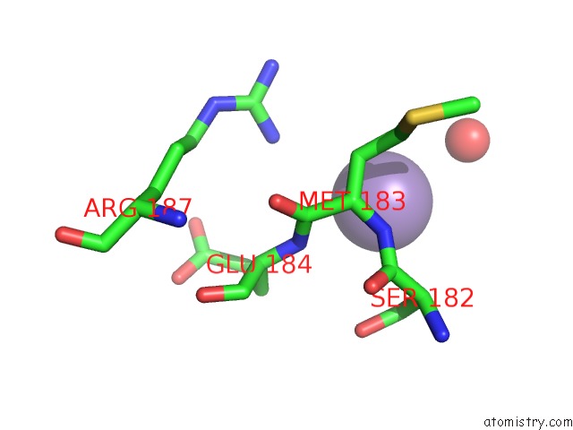
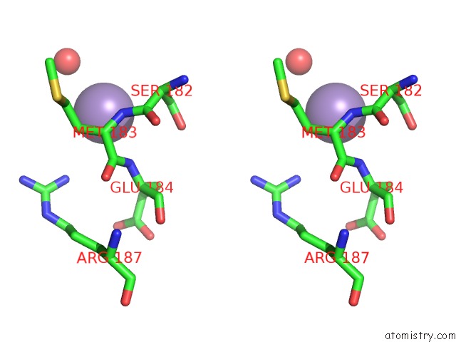
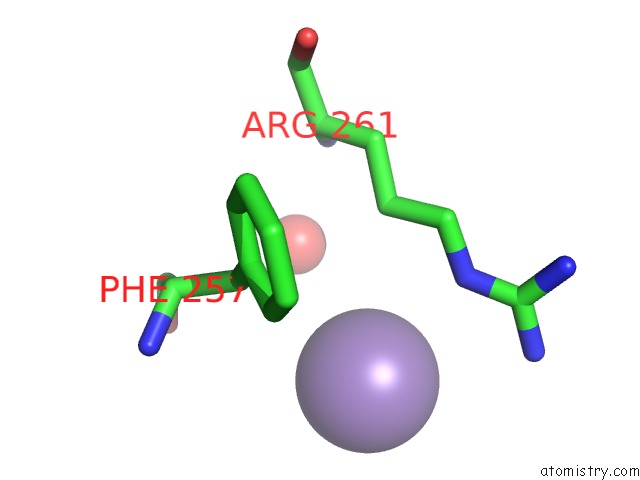
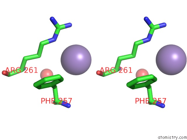
Manganese binding site 1 out of 8 in 2bcd
Go back to
Manganese binding site 1 out
of 8 in the X-Ray Crystal Structure of Protein Phosphatase-1 with the Marine Toxin Motuporin Bound
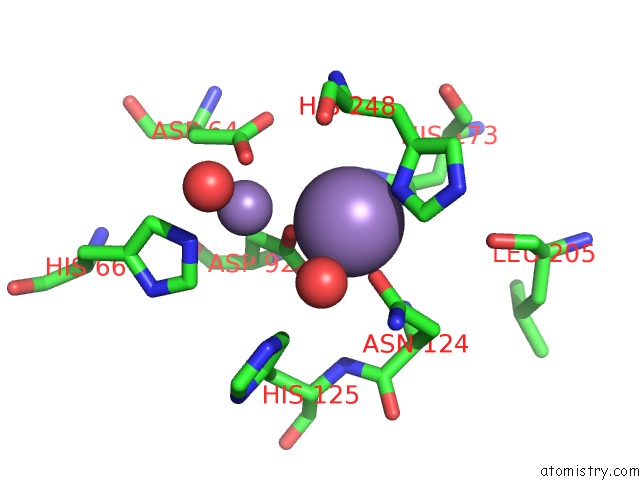
Mono view
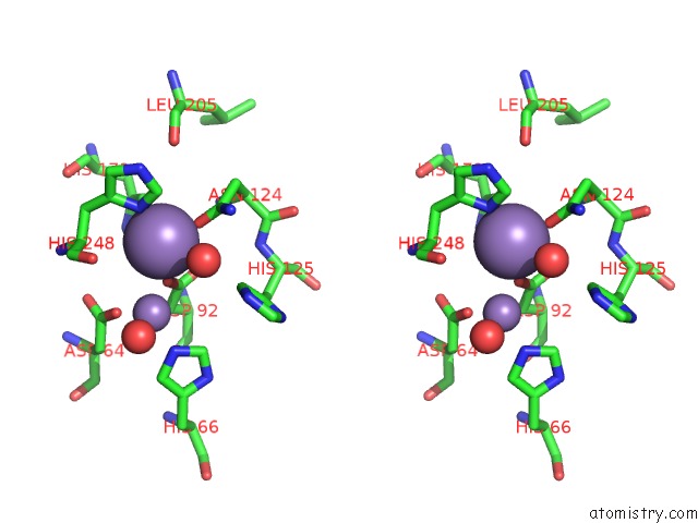
Stereo pair view
Mono view
Stereo pair view
A full contact list of Manganese with other atoms in the Mn binding
site number 1 of X-Ray Crystal Structure of Protein Phosphatase-1 with the Marine Toxin Motuporin Bound within 5.0Å range:
|
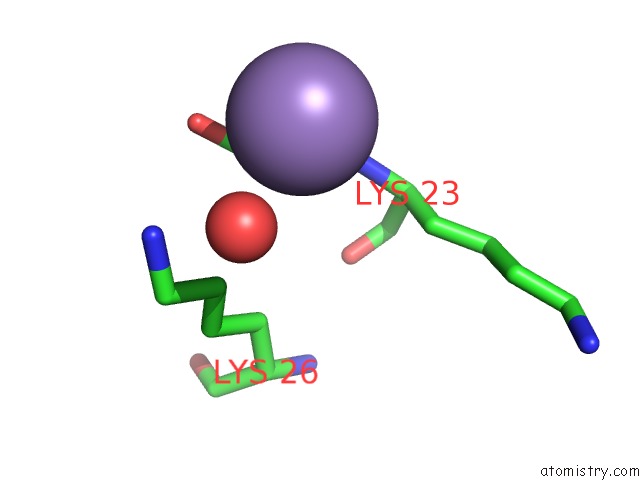
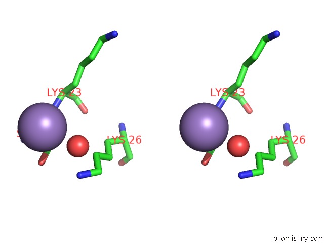
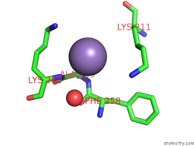
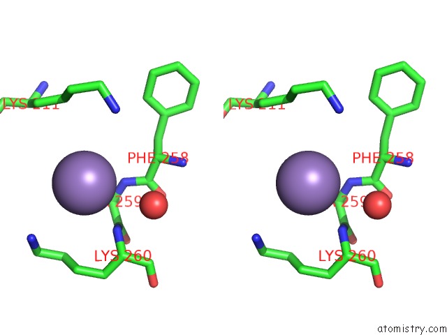
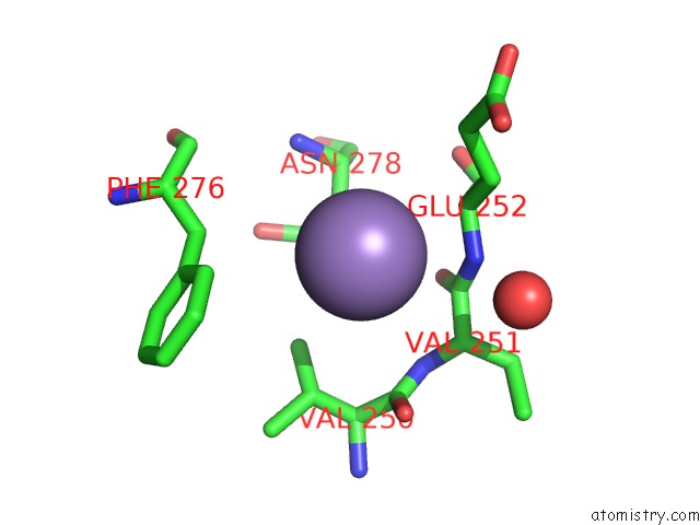
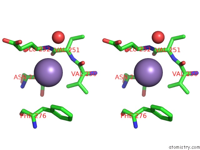
Manganese binding site 2 out of 8 in 2bcd
Go back to
Manganese binding site 2 out
of 8 in the X-Ray Crystal Structure of Protein Phosphatase-1 with the Marine Toxin Motuporin Bound
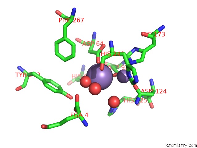
Mono view
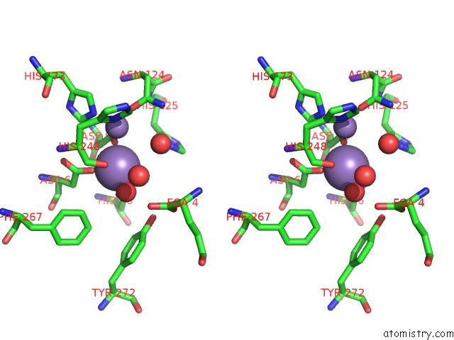
Stereo pair view
Mono view
Stereo pair view
A full contact list of Manganese with other atoms in the Mn binding
site number 2 of X-Ray Crystal Structure of Protein Phosphatase-1 with the Marine Toxin Motuporin Bound within 5.0Å range:
|
Manganese binding site 3 out of 8 in 2bcd
Go back to
Manganese binding site 3 out
of 8 in the X-Ray Crystal Structure of Protein Phosphatase-1 with the Marine Toxin Motuporin Bound
Mono view
Stereo pair view
Mono view
Stereo pair view
A full contact list of Manganese with other atoms in the Mn binding
site number 3 of X-Ray Crystal Structure of Protein Phosphatase-1 with the Marine Toxin Motuporin Bound within 5.0Å range:
|
Manganese binding site 4 out of 8 in 2bcd
Go back to
Manganese binding site 4 out
of 8 in the X-Ray Crystal Structure of Protein Phosphatase-1 with the Marine Toxin Motuporin Bound
Mono view
Stereo pair view
Mono view
Stereo pair view
A full contact list of Manganese with other atoms in the Mn binding
site number 4 of X-Ray Crystal Structure of Protein Phosphatase-1 with the Marine Toxin Motuporin Bound within 5.0Å range:
|
Manganese binding site 5 out of 8 in 2bcd
Go back to
Manganese binding site 5 out
of 8 in the X-Ray Crystal Structure of Protein Phosphatase-1 with the Marine Toxin Motuporin Bound
Mono view
Stereo pair view
Mono view
Stereo pair view
A full contact list of Manganese with other atoms in the Mn binding
site number 5 of X-Ray Crystal Structure of Protein Phosphatase-1 with the Marine Toxin Motuporin Bound within 5.0Å range:
|
Manganese binding site 6 out of 8 in 2bcd
Go back to
Manganese binding site 6 out
of 8 in the X-Ray Crystal Structure of Protein Phosphatase-1 with the Marine Toxin Motuporin Bound
Mono view
Stereo pair view
Mono view
Stereo pair view
A full contact list of Manganese with other atoms in the Mn binding
site number 6 of X-Ray Crystal Structure of Protein Phosphatase-1 with the Marine Toxin Motuporin Bound within 5.0Å range:
|
Manganese binding site 7 out of 8 in 2bcd
Go back to
Manganese binding site 7 out
of 8 in the X-Ray Crystal Structure of Protein Phosphatase-1 with the Marine Toxin Motuporin Bound
Mono view
Stereo pair view
Mono view
Stereo pair view
A full contact list of Manganese with other atoms in the Mn binding
site number 7 of X-Ray Crystal Structure of Protein Phosphatase-1 with the Marine Toxin Motuporin Bound within 5.0Å range:
|
Manganese binding site 8 out of 8 in 2bcd
Go back to
Manganese binding site 8 out
of 8 in the X-Ray Crystal Structure of Protein Phosphatase-1 with the Marine Toxin Motuporin Bound
Mono view
Stereo pair view
Mono view
Stereo pair view
A full contact list of Manganese with other atoms in the Mn binding
site number 8 of X-Ray Crystal Structure of Protein Phosphatase-1 with the Marine Toxin Motuporin Bound within 5.0Å range:
|
Reference:
J.T.Maynes,
H.A.Luu,
M.M.Cherney,
R.J.Andersen,
D.Williams,
C.F.Holmes,
M.N.James.
Crystal Structures of Protein Phosphatase-1 Bound to Motuporin and Dihydromicrocystin-La: Elucidation of the Mechanism of Enzyme Inhibition By Cyanobacterial Toxins. J.Mol.Biol. V. 356 111 2006.
ISSN: ISSN 0022-2836
PubMed: 16343532
DOI: 10.1016/J.JMB.2005.11.019
Page generated: Sat Oct 5 13:32:54 2024
ISSN: ISSN 0022-2836
PubMed: 16343532
DOI: 10.1016/J.JMB.2005.11.019
Last articles
K in 7LJ5K in 7LJ4
K in 7LFT
K in 7LC3
K in 7LFQ
K in 7LC6
K in 7L4G
K in 7L0Z
K in 7KZ0
K in 7L1A