Manganese »
PDB 2ayl-2ce4 »
2b7o »
Manganese in PDB 2b7o: The Structure of 3-Deoxy-D-Arabino-Heptulosonate 7-Phosphate Synthase From Mycobacterium Tuberculosis
Enzymatic activity of The Structure of 3-Deoxy-D-Arabino-Heptulosonate 7-Phosphate Synthase From Mycobacterium Tuberculosis
All present enzymatic activity of The Structure of 3-Deoxy-D-Arabino-Heptulosonate 7-Phosphate Synthase From Mycobacterium Tuberculosis:
2.5.1.54;
2.5.1.54;
Protein crystallography data
The structure of The Structure of 3-Deoxy-D-Arabino-Heptulosonate 7-Phosphate Synthase From Mycobacterium Tuberculosis, PDB code: 2b7o
was solved by
C.J.Webby,
H.M.Baker,
J.S.Lott,
E.N.Baker,
E.J.Parker,
Mycobacteriumtuberculosis Structural Proteomics Project (Xmtb),
with X-Ray Crystallography technique. A brief refinement statistics is given in the table below:
| Resolution Low / High (Å) | 47.04 / 2.30 |
| Space group | P 32 2 1 |
| Cell size a, b, c (Å), α, β, γ (°) | 204.085, 204.085, 66.230, 90.00, 90.00, 120.00 |
| R / Rfree (%) | 18.8 / 22.4 |
Manganese Binding Sites:
The binding sites of Manganese atom in the The Structure of 3-Deoxy-D-Arabino-Heptulosonate 7-Phosphate Synthase From Mycobacterium Tuberculosis
(pdb code 2b7o). This binding sites where shown within
5.0 Angstroms radius around Manganese atom.
In total 2 binding sites of Manganese where determined in the The Structure of 3-Deoxy-D-Arabino-Heptulosonate 7-Phosphate Synthase From Mycobacterium Tuberculosis, PDB code: 2b7o:
Jump to Manganese binding site number: 1; 2;
In total 2 binding sites of Manganese where determined in the The Structure of 3-Deoxy-D-Arabino-Heptulosonate 7-Phosphate Synthase From Mycobacterium Tuberculosis, PDB code: 2b7o:
Jump to Manganese binding site number: 1; 2;
Manganese binding site 1 out of 2 in 2b7o
Go back to
Manganese binding site 1 out
of 2 in the The Structure of 3-Deoxy-D-Arabino-Heptulosonate 7-Phosphate Synthase From Mycobacterium Tuberculosis
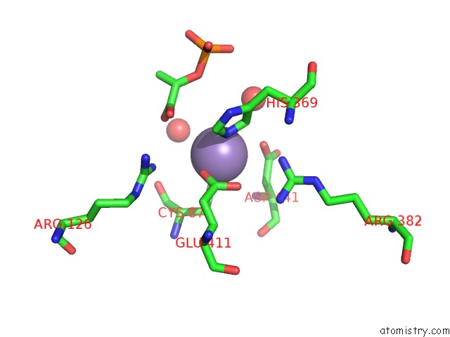
Mono view
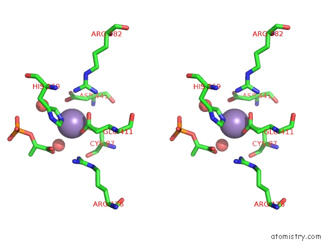
Stereo pair view
Mono view
Stereo pair view
A full contact list of Manganese with other atoms in the Mn binding
site number 1 of The Structure of 3-Deoxy-D-Arabino-Heptulosonate 7-Phosphate Synthase From Mycobacterium Tuberculosis within 5.0Å range:
|
Manganese binding site 2 out of 2 in 2b7o
Go back to
Manganese binding site 2 out
of 2 in the The Structure of 3-Deoxy-D-Arabino-Heptulosonate 7-Phosphate Synthase From Mycobacterium Tuberculosis
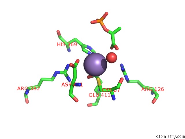
Mono view
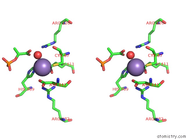
Stereo pair view
Mono view
Stereo pair view
A full contact list of Manganese with other atoms in the Mn binding
site number 2 of The Structure of 3-Deoxy-D-Arabino-Heptulosonate 7-Phosphate Synthase From Mycobacterium Tuberculosis within 5.0Å range:
|
Reference:
C.J.Webby,
H.M.Baker,
J.S.Lott,
E.N.Baker,
E.J.Parker.
The Structure of 3-Deoxy-D-Arabino-Heptulosonate 7-Phosphate Synthase From Mycobacterium Tuberculosis Reveals A Common Catalytic Scaffold and Ancestry For Type I and Type II Enzymes J.Mol.Biol. V. 354 927 2005.
ISSN: ISSN 0022-2836
PubMed: 16288916
DOI: 10.1016/J.JMB.2005.09.093
Page generated: Sat Oct 5 13:32:15 2024
ISSN: ISSN 0022-2836
PubMed: 16288916
DOI: 10.1016/J.JMB.2005.09.093
Last articles
K in 6CK5K in 6CGP
K in 6CF1
K in 6CI0
K in 6C9X
K in 6C65
K in 6C64
K in 6C9U
K in 6C63
K in 6C0Y