Manganese »
PDB 1yw7-2a2t »
1zop »
Manganese in PDB 1zop: CD11A I-Domain with Bound Magnesium Ion
Protein crystallography data
The structure of CD11A I-Domain with Bound Magnesium Ion, PDB code: 1zop
was solved by
D.J.Leahy,
A.Qu,
with X-Ray Crystallography technique. A brief refinement statistics is given in the table below:
| Resolution Low / High (Å) | 30.00 / 2.00 |
| Space group | P 21 21 2 |
| Cell size a, b, c (Å), α, β, γ (°) | 76.160, 78.630, 66.370, 90.00, 90.00, 90.00 |
| R / Rfree (%) | 19.7 / 26.3 |
Other elements in 1zop:
The structure of CD11A I-Domain with Bound Magnesium Ion also contains other interesting chemical elements:
| Chlorine | (Cl) | 2 atoms |
Manganese Binding Sites:
The binding sites of Manganese atom in the CD11A I-Domain with Bound Magnesium Ion
(pdb code 1zop). This binding sites where shown within
5.0 Angstroms radius around Manganese atom.
In total 2 binding sites of Manganese where determined in the CD11A I-Domain with Bound Magnesium Ion, PDB code: 1zop:
Jump to Manganese binding site number: 1; 2;
In total 2 binding sites of Manganese where determined in the CD11A I-Domain with Bound Magnesium Ion, PDB code: 1zop:
Jump to Manganese binding site number: 1; 2;
Manganese binding site 1 out of 2 in 1zop
Go back to
Manganese binding site 1 out
of 2 in the CD11A I-Domain with Bound Magnesium Ion
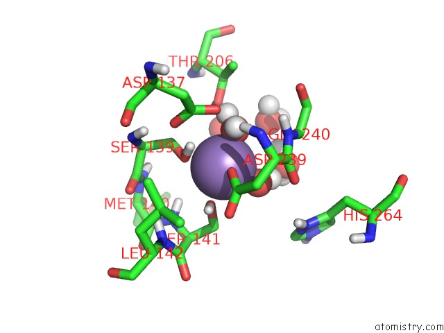
Mono view
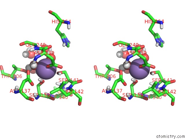
Stereo pair view
Mono view
Stereo pair view
A full contact list of Manganese with other atoms in the Mn binding
site number 1 of CD11A I-Domain with Bound Magnesium Ion within 5.0Å range:
|
Manganese binding site 2 out of 2 in 1zop
Go back to
Manganese binding site 2 out
of 2 in the CD11A I-Domain with Bound Magnesium Ion
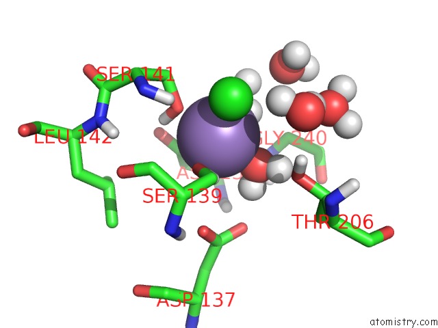
Mono view
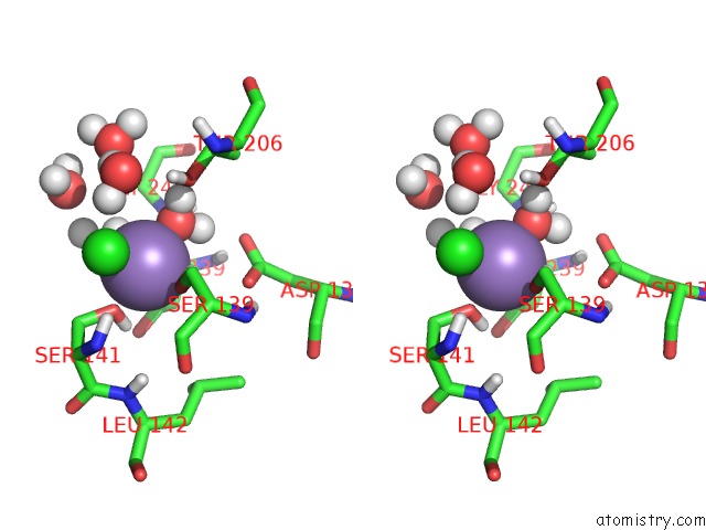
Stereo pair view
Mono view
Stereo pair view
A full contact list of Manganese with other atoms in the Mn binding
site number 2 of CD11A I-Domain with Bound Magnesium Ion within 5.0Å range:
|
Reference:
A.Qu,
D.J.Leahy.
The Role of the Divalent Cation in the Structure of the I Domain From the CD11A/CD18 Integrin. Structure V. 4 931 1996.
ISSN: ISSN 0969-2126
PubMed: 8805579
DOI: 10.1016/S0969-2126(96)00100-1
Page generated: Sat Oct 5 13:18:06 2024
ISSN: ISSN 0969-2126
PubMed: 8805579
DOI: 10.1016/S0969-2126(96)00100-1
Last articles
Fe in 2YXOFe in 2YRS
Fe in 2YXC
Fe in 2YNM
Fe in 2YVJ
Fe in 2YP1
Fe in 2YU2
Fe in 2YU1
Fe in 2YQB
Fe in 2YOO