Manganese »
PDB 1yw7-2a2t »
1z2w »
Manganese in PDB 1z2w: Crystal Structure of Mouse VPS29 Complexed with MN2+
Protein crystallography data
The structure of Crystal Structure of Mouse VPS29 Complexed with MN2+, PDB code: 1z2w
was solved by
B.M.Collins,
C.F.Skinner,
P.J.Watson,
M.N.J.Seaman,
D.J.Owen,
with X-Ray Crystallography technique. A brief refinement statistics is given in the table below:
| Resolution Low / High (Å) | 20.00 / 2.00 |
| Space group | P 1 21 1 |
| Cell size a, b, c (Å), α, β, γ (°) | 55.914, 68.953, 60.519, 90.00, 106.65, 90.00 |
| R / Rfree (%) | 17.6 / 23.7 |
Manganese Binding Sites:
The binding sites of Manganese atom in the Crystal Structure of Mouse VPS29 Complexed with MN2+
(pdb code 1z2w). This binding sites where shown within
5.0 Angstroms radius around Manganese atom.
In total 4 binding sites of Manganese where determined in the Crystal Structure of Mouse VPS29 Complexed with MN2+, PDB code: 1z2w:
Jump to Manganese binding site number: 1; 2; 3; 4;
In total 4 binding sites of Manganese where determined in the Crystal Structure of Mouse VPS29 Complexed with MN2+, PDB code: 1z2w:
Jump to Manganese binding site number: 1; 2; 3; 4;
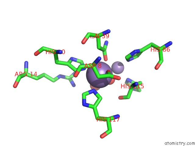
Manganese binding site 1 out of 4 in 1z2w
Go back to
Manganese binding site 1 out
of 4 in the Crystal Structure of Mouse VPS29 Complexed with MN2+
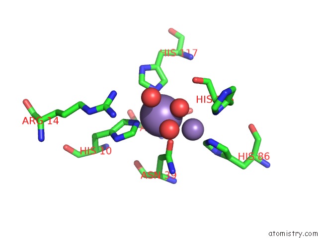
Mono view
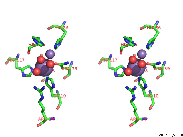
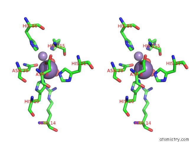
Stereo pair view
Mono view
Stereo pair view
A full contact list of Manganese with other atoms in the Mn binding
site number 1 of Crystal Structure of Mouse VPS29 Complexed with MN2+ within 5.0Å range:
|
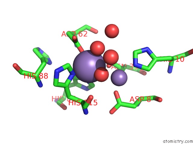
Manganese binding site 2 out of 4 in 1z2w
Go back to
Manganese binding site 2 out
of 4 in the Crystal Structure of Mouse VPS29 Complexed with MN2+
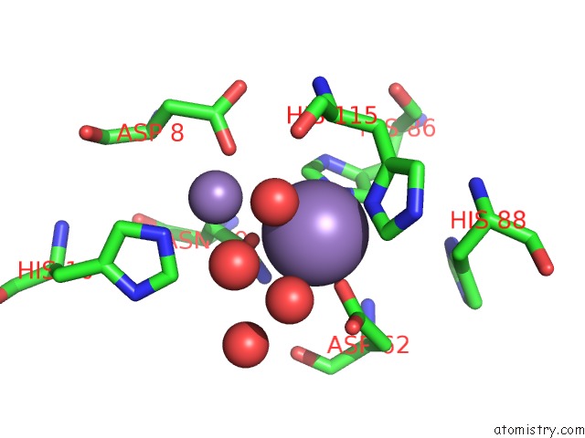
Mono view
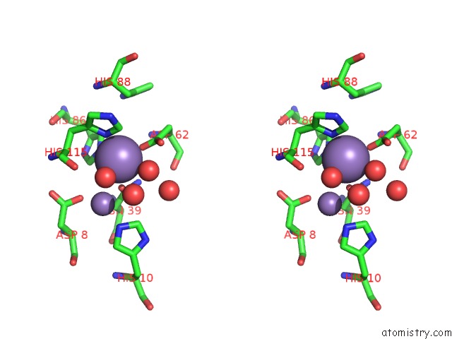
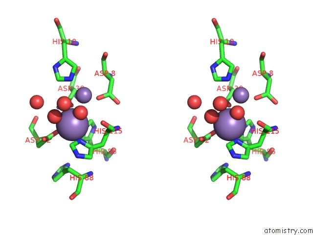
Stereo pair view
Mono view
Stereo pair view
A full contact list of Manganese with other atoms in the Mn binding
site number 2 of Crystal Structure of Mouse VPS29 Complexed with MN2+ within 5.0Å range:
|
Manganese binding site 3 out of 4 in 1z2w
Go back to
Manganese binding site 3 out
of 4 in the Crystal Structure of Mouse VPS29 Complexed with MN2+
Mono view
Stereo pair view
Mono view
Stereo pair view
A full contact list of Manganese with other atoms in the Mn binding
site number 3 of Crystal Structure of Mouse VPS29 Complexed with MN2+ within 5.0Å range:
|
Manganese binding site 4 out of 4 in 1z2w
Go back to
Manganese binding site 4 out
of 4 in the Crystal Structure of Mouse VPS29 Complexed with MN2+
Mono view
Stereo pair view
Mono view
Stereo pair view
A full contact list of Manganese with other atoms in the Mn binding
site number 4 of Crystal Structure of Mouse VPS29 Complexed with MN2+ within 5.0Å range:
|
Reference:
B.M.Collins,
C.F.Skinner,
P.J.Watson,
M.N.J.Seaman,
D.J.Owen.
VPS29 Has A Phosphoesterase Fold That Acts As A Protein Interaction Scaffold For Retromer Assembly Nat.Struct.Mol.Biol. V. 12 594 2005.
ISSN: ISSN 1545-9993
PubMed: 15965486
DOI: 10.1038/NSMB954
Page generated: Sat Oct 5 13:15:51 2024
ISSN: ISSN 1545-9993
PubMed: 15965486
DOI: 10.1038/NSMB954
Last articles
Mg in 3U2NMg in 3U2K
Mg in 3U2E
Mg in 3U2Q
Mg in 3U2D
Mg in 3TZR
Mg in 3U0U
Mg in 3U13
Mg in 3U0O
Mg in 3U08