Manganese »
PDB 1xif-1ytm »
1yny »
Manganese in PDB 1yny: Molecular Structure of D-Hydantoinase From A Bacillus Sp. AR9: Evidence For Mercury Inhibition
Enzymatic activity of Molecular Structure of D-Hydantoinase From A Bacillus Sp. AR9: Evidence For Mercury Inhibition
All present enzymatic activity of Molecular Structure of D-Hydantoinase From A Bacillus Sp. AR9: Evidence For Mercury Inhibition:
3.5.2.2;
3.5.2.2;
Protein crystallography data
The structure of Molecular Structure of D-Hydantoinase From A Bacillus Sp. AR9: Evidence For Mercury Inhibition, PDB code: 1yny
was solved by
K.V.Radha Kishan,
R.M.Vohra,
K.Ganeshan,
V.Agrawal,
V.M.Sharma,
R.Sharma,
with X-Ray Crystallography technique. A brief refinement statistics is given in the table below:
| Resolution Low / High (Å) | 50.00 / 2.30 |
| Space group | P 64 |
| Cell size a, b, c (Å), α, β, γ (°) | 129.540, 129.540, 102.850, 90.00, 90.00, 120.00 |
| R / Rfree (%) | 19.9 / 24.3 |
Manganese Binding Sites:
The binding sites of Manganese atom in the Molecular Structure of D-Hydantoinase From A Bacillus Sp. AR9: Evidence For Mercury Inhibition
(pdb code 1yny). This binding sites where shown within
5.0 Angstroms radius around Manganese atom.
In total 4 binding sites of Manganese where determined in the Molecular Structure of D-Hydantoinase From A Bacillus Sp. AR9: Evidence For Mercury Inhibition, PDB code: 1yny:
Jump to Manganese binding site number: 1; 2; 3; 4;
In total 4 binding sites of Manganese where determined in the Molecular Structure of D-Hydantoinase From A Bacillus Sp. AR9: Evidence For Mercury Inhibition, PDB code: 1yny:
Jump to Manganese binding site number: 1; 2; 3; 4;
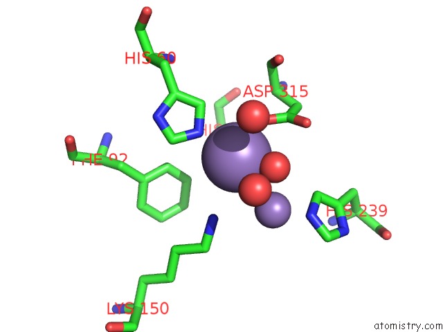
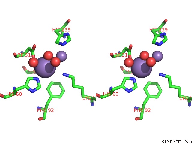
Manganese binding site 1 out of 4 in 1yny
Go back to
Manganese binding site 1 out
of 4 in the Molecular Structure of D-Hydantoinase From A Bacillus Sp. AR9: Evidence For Mercury Inhibition
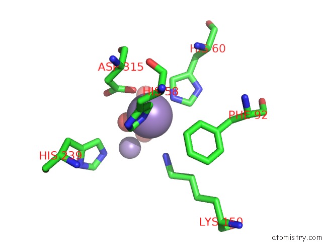
Mono view
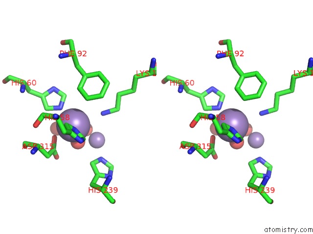
Stereo pair view
Mono view
Stereo pair view
A full contact list of Manganese with other atoms in the Mn binding
site number 1 of Molecular Structure of D-Hydantoinase From A Bacillus Sp. AR9: Evidence For Mercury Inhibition within 5.0Å range:
|
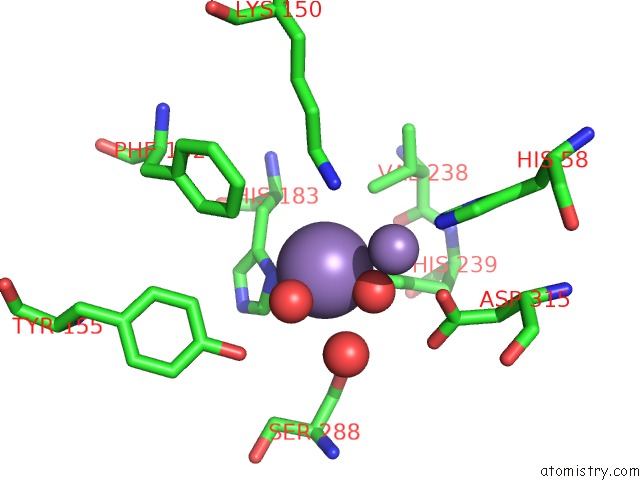
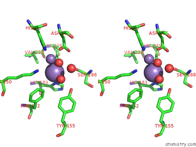
Manganese binding site 2 out of 4 in 1yny
Go back to
Manganese binding site 2 out
of 4 in the Molecular Structure of D-Hydantoinase From A Bacillus Sp. AR9: Evidence For Mercury Inhibition
Mono view
Stereo pair view
Mono view
Stereo pair view
A full contact list of Manganese with other atoms in the Mn binding
site number 2 of Molecular Structure of D-Hydantoinase From A Bacillus Sp. AR9: Evidence For Mercury Inhibition within 5.0Å range:
|
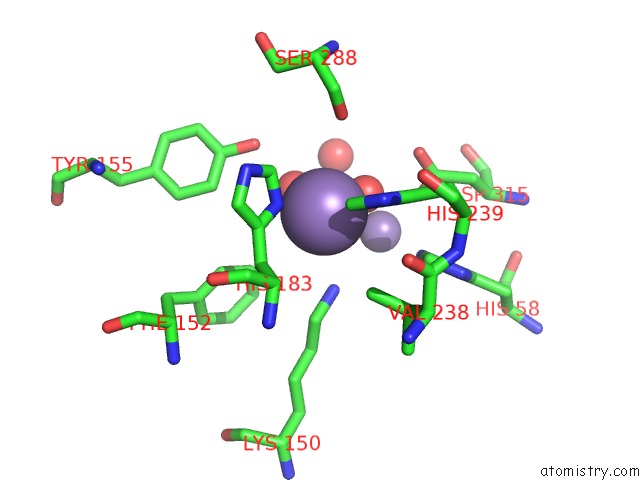
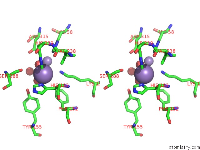
Manganese binding site 3 out of 4 in 1yny
Go back to
Manganese binding site 3 out
of 4 in the Molecular Structure of D-Hydantoinase From A Bacillus Sp. AR9: Evidence For Mercury Inhibition
Mono view
Stereo pair view
Mono view
Stereo pair view
A full contact list of Manganese with other atoms in the Mn binding
site number 3 of Molecular Structure of D-Hydantoinase From A Bacillus Sp. AR9: Evidence For Mercury Inhibition within 5.0Å range:
|
Manganese binding site 4 out of 4 in 1yny
Go back to
Manganese binding site 4 out
of 4 in the Molecular Structure of D-Hydantoinase From A Bacillus Sp. AR9: Evidence For Mercury Inhibition
Mono view
Stereo pair view
Mono view
Stereo pair view
A full contact list of Manganese with other atoms in the Mn binding
site number 4 of Molecular Structure of D-Hydantoinase From A Bacillus Sp. AR9: Evidence For Mercury Inhibition within 5.0Å range:
|
Reference:
K.V.Radha Kishan,
R.M.Vohra,
K.Ganesan,
V.Agrawal,
V.M.Sharma,
R.Sharma.
Molecular Structure of D-Hydantoinase From Bacillus Sp. AR9: Evidence For Mercury Inhibition. J.Mol.Biol. V. 347 95 2005.
ISSN: ISSN 0022-2836
PubMed: 15733920
DOI: 10.1016/J.JMB.2005.01.025
Page generated: Sat Oct 5 13:13:17 2024
ISSN: ISSN 0022-2836
PubMed: 15733920
DOI: 10.1016/J.JMB.2005.01.025
Last articles
K in 9FYEK in 9FT7
K in 9FQ1
K in 9FM9
K in 9EX3
K in 9F90
K in 9ES6
K in 9EWD
K in 9ETN
K in 9ESI