Manganese »
PDB 1xif-1ytm »
1ybu »
Manganese in PDB 1ybu: Mycobacterium Tuberculosis Adenylyl Cyclase RV1900C Chd, in Complex with A Substrate Analog.
Enzymatic activity of Mycobacterium Tuberculosis Adenylyl Cyclase RV1900C Chd, in Complex with A Substrate Analog.
All present enzymatic activity of Mycobacterium Tuberculosis Adenylyl Cyclase RV1900C Chd, in Complex with A Substrate Analog.:
4.6.1.1;
4.6.1.1;
Protein crystallography data
The structure of Mycobacterium Tuberculosis Adenylyl Cyclase RV1900C Chd, in Complex with A Substrate Analog., PDB code: 1ybu
was solved by
S.C.Sinha,
M.Wetterer,
S.R.Sprang,
J.E.Schultz,
J.U.Linder,
with X-Ray Crystallography technique. A brief refinement statistics is given in the table below:
| Resolution Low / High (Å) | 31.94 / 2.40 |
| Space group | P 1 2 1 |
| Cell size a, b, c (Å), α, β, γ (°) | 91.067, 48.923, 68.198, 90.00, 90.00, 90.00 |
| R / Rfree (%) | 23.3 / 26.2 |
Manganese Binding Sites:
The binding sites of Manganese atom in the Mycobacterium Tuberculosis Adenylyl Cyclase RV1900C Chd, in Complex with A Substrate Analog.
(pdb code 1ybu). This binding sites where shown within
5.0 Angstroms radius around Manganese atom.
In total 2 binding sites of Manganese where determined in the Mycobacterium Tuberculosis Adenylyl Cyclase RV1900C Chd, in Complex with A Substrate Analog., PDB code: 1ybu:
Jump to Manganese binding site number: 1; 2;
In total 2 binding sites of Manganese where determined in the Mycobacterium Tuberculosis Adenylyl Cyclase RV1900C Chd, in Complex with A Substrate Analog., PDB code: 1ybu:
Jump to Manganese binding site number: 1; 2;
Manganese binding site 1 out of 2 in 1ybu
Go back to
Manganese binding site 1 out
of 2 in the Mycobacterium Tuberculosis Adenylyl Cyclase RV1900C Chd, in Complex with A Substrate Analog.
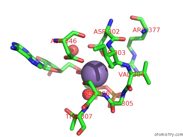
Mono view
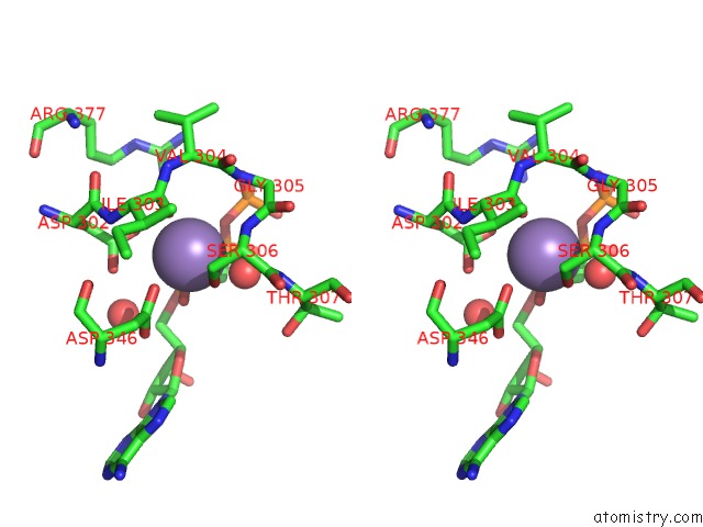
Stereo pair view
Mono view
Stereo pair view
A full contact list of Manganese with other atoms in the Mn binding
site number 1 of Mycobacterium Tuberculosis Adenylyl Cyclase RV1900C Chd, in Complex with A Substrate Analog. within 5.0Å range:
|
Manganese binding site 2 out of 2 in 1ybu
Go back to
Manganese binding site 2 out
of 2 in the Mycobacterium Tuberculosis Adenylyl Cyclase RV1900C Chd, in Complex with A Substrate Analog.
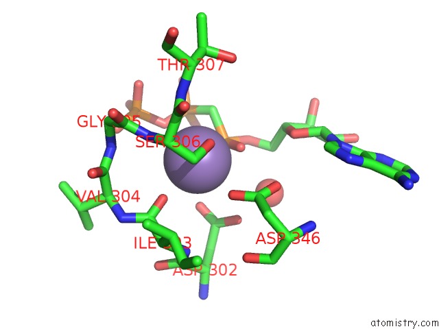
Mono view
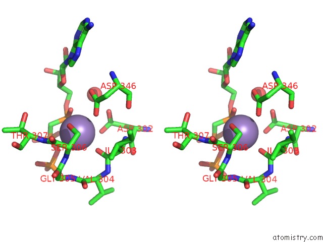
Stereo pair view
Mono view
Stereo pair view
A full contact list of Manganese with other atoms in the Mn binding
site number 2 of Mycobacterium Tuberculosis Adenylyl Cyclase RV1900C Chd, in Complex with A Substrate Analog. within 5.0Å range:
|
Reference:
S.C.Sinha,
M.Wetterer,
S.R.Sprang,
J.E.Schultz,
J.U.Linder.
Origin of Asymmetry in Adenylyl Cyclases: Structures of Mycobacterium Tuberculosis RV1900C. Embo J. V. 24 663 2005.
ISSN: ISSN 0261-4189
PubMed: 15678099
DOI: 10.1038/SJ.EMBOJ.7600573
Page generated: Sat Oct 5 13:10:26 2024
ISSN: ISSN 0261-4189
PubMed: 15678099
DOI: 10.1038/SJ.EMBOJ.7600573
Last articles
K in 9EX3K in 9F90
K in 9ES6
K in 9EWD
K in 9ETN
K in 9ESI
K in 9ESH
K in 9ES4
K in 9ES5
K in 9ES2