Manganese »
PDB 1xif-1ytm »
1xmf »
Manganese in PDB 1xmf: Structure of Mn(II)-Soaked Apo Methane Monooxygenase Hydroxylase Crystals From M. Capsulatus (Bath)
Enzymatic activity of Structure of Mn(II)-Soaked Apo Methane Monooxygenase Hydroxylase Crystals From M. Capsulatus (Bath)
All present enzymatic activity of Structure of Mn(II)-Soaked Apo Methane Monooxygenase Hydroxylase Crystals From M. Capsulatus (Bath):
1.14.13.25;
1.14.13.25;
Protein crystallography data
The structure of Structure of Mn(II)-Soaked Apo Methane Monooxygenase Hydroxylase Crystals From M. Capsulatus (Bath), PDB code: 1xmf
was solved by
M.H.Sazinsky,
M.Merkx,
E.Cadieux,
S.Tang,
S.J.Lippard,
with X-Ray Crystallography technique. A brief refinement statistics is given in the table below:
| Resolution Low / High (Å) | 28.76 / 2.32 |
| Space group | P 21 21 21 |
| Cell size a, b, c (Å), α, β, γ (°) | 70.820, 171.679, 220.275, 90.00, 90.00, 90.00 |
| R / Rfree (%) | 21.9 / 26.3 |
Other elements in 1xmf:
The structure of Structure of Mn(II)-Soaked Apo Methane Monooxygenase Hydroxylase Crystals From M. Capsulatus (Bath) also contains other interesting chemical elements:
| Calcium | (Ca) | 4 atoms |
Manganese Binding Sites:
The binding sites of Manganese atom in the Structure of Mn(II)-Soaked Apo Methane Monooxygenase Hydroxylase Crystals From M. Capsulatus (Bath)
(pdb code 1xmf). This binding sites where shown within
5.0 Angstroms radius around Manganese atom.
In total 4 binding sites of Manganese where determined in the Structure of Mn(II)-Soaked Apo Methane Monooxygenase Hydroxylase Crystals From M. Capsulatus (Bath), PDB code: 1xmf:
Jump to Manganese binding site number: 1; 2; 3; 4;
In total 4 binding sites of Manganese where determined in the Structure of Mn(II)-Soaked Apo Methane Monooxygenase Hydroxylase Crystals From M. Capsulatus (Bath), PDB code: 1xmf:
Jump to Manganese binding site number: 1; 2; 3; 4;
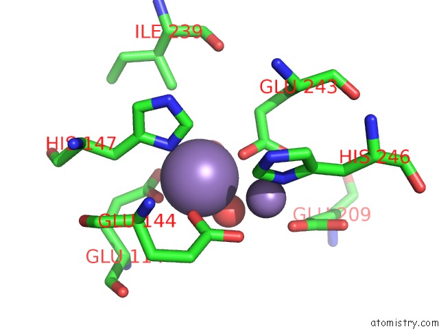
Manganese binding site 1 out of 4 in 1xmf
Go back to
Manganese binding site 1 out
of 4 in the Structure of Mn(II)-Soaked Apo Methane Monooxygenase Hydroxylase Crystals From M. Capsulatus (Bath)
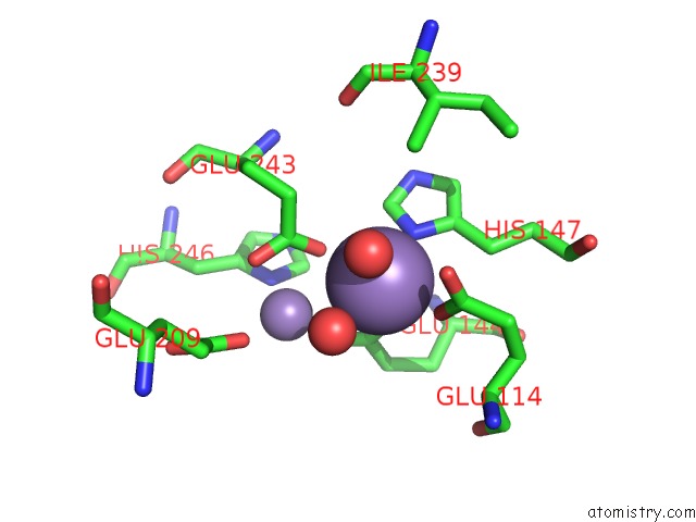
Mono view
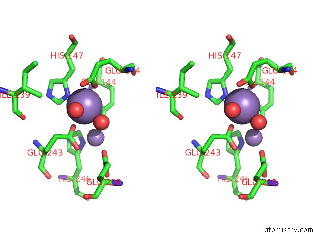
Stereo pair view
Mono view
Stereo pair view
A full contact list of Manganese with other atoms in the Mn binding
site number 1 of Structure of Mn(II)-Soaked Apo Methane Monooxygenase Hydroxylase Crystals From M. Capsulatus (Bath) within 5.0Å range:
|
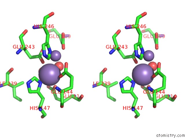
Manganese binding site 2 out of 4 in 1xmf
Go back to
Manganese binding site 2 out
of 4 in the Structure of Mn(II)-Soaked Apo Methane Monooxygenase Hydroxylase Crystals From M. Capsulatus (Bath)
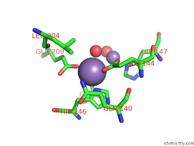
Mono view
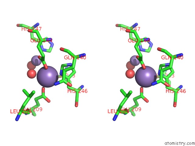
Stereo pair view
Mono view
Stereo pair view
A full contact list of Manganese with other atoms in the Mn binding
site number 2 of Structure of Mn(II)-Soaked Apo Methane Monooxygenase Hydroxylase Crystals From M. Capsulatus (Bath) within 5.0Å range:
|
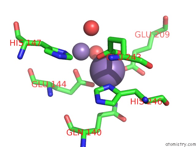
Manganese binding site 3 out of 4 in 1xmf
Go back to
Manganese binding site 3 out
of 4 in the Structure of Mn(II)-Soaked Apo Methane Monooxygenase Hydroxylase Crystals From M. Capsulatus (Bath)
Mono view
Stereo pair view
Mono view
Stereo pair view
A full contact list of Manganese with other atoms in the Mn binding
site number 3 of Structure of Mn(II)-Soaked Apo Methane Monooxygenase Hydroxylase Crystals From M. Capsulatus (Bath) within 5.0Å range:
|
Manganese binding site 4 out of 4 in 1xmf
Go back to
Manganese binding site 4 out
of 4 in the Structure of Mn(II)-Soaked Apo Methane Monooxygenase Hydroxylase Crystals From M. Capsulatus (Bath)
Mono view
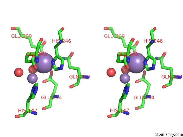
Stereo pair view
Mono view
Stereo pair view
A full contact list of Manganese with other atoms in the Mn binding
site number 4 of Structure of Mn(II)-Soaked Apo Methane Monooxygenase Hydroxylase Crystals From M. Capsulatus (Bath) within 5.0Å range:
|
Reference:
M.H.Sazinsky,
M.Merkx,
E.Cadieux,
S.Tang,
S.J.Lippard.
Preparation and X-Ray Structures of Metal-Free, Dicobalt and Dimanganese Forms of Soluble Methane Monooxygenase Hydroxylase From Methylococcus Capsulatus (Bath) Biochemistry V. 43 16263 2004.
ISSN: ISSN 0006-2960
PubMed: 15610020
DOI: 10.1021/BI048140Z
Page generated: Sat Oct 5 13:07:29 2024
ISSN: ISSN 0006-2960
PubMed: 15610020
DOI: 10.1021/BI048140Z
Last articles
K in 9FYEK in 9FT7
K in 9FQ1
K in 9FM9
K in 9EX3
K in 9F90
K in 9ES6
K in 9EWD
K in 9ETN
K in 9ESI