Manganese »
PDB 1pj4-1r1o »
1qb4 »
Manganese in PDB 1qb4: Crystal Structure of Mn(2+)-Bound Phosphoenolpyruvate Carboxylase
Enzymatic activity of Crystal Structure of Mn(2+)-Bound Phosphoenolpyruvate Carboxylase
All present enzymatic activity of Crystal Structure of Mn(2+)-Bound Phosphoenolpyruvate Carboxylase:
4.1.1.31;
4.1.1.31;
Protein crystallography data
The structure of Crystal Structure of Mn(2+)-Bound Phosphoenolpyruvate Carboxylase, PDB code: 1qb4
was solved by
H.Matsumura,
M.Terada,
S.Shirakata,
T.Inoue,
T.Yoshinaga,
K.Izui,
Y.Kai,
with X-Ray Crystallography technique. A brief refinement statistics is given in the table below:
| Resolution Low / High (Å) | 20.00 / 2.60 |
| Space group | I 2 2 2 |
| Cell size a, b, c (Å), α, β, γ (°) | 117.750, 248.410, 83.470, 90.00, 90.00, 90.00 |
| R / Rfree (%) | 22.3 / 26.1 |
Manganese Binding Sites:
The binding sites of Manganese atom in the Crystal Structure of Mn(2+)-Bound Phosphoenolpyruvate Carboxylase
(pdb code 1qb4). This binding sites where shown within
5.0 Angstroms radius around Manganese atom.
In total only one binding site of Manganese was determined in the Crystal Structure of Mn(2+)-Bound Phosphoenolpyruvate Carboxylase, PDB code: 1qb4:
In total only one binding site of Manganese was determined in the Crystal Structure of Mn(2+)-Bound Phosphoenolpyruvate Carboxylase, PDB code: 1qb4:
Manganese binding site 1 out of 1 in 1qb4
Go back to
Manganese binding site 1 out
of 1 in the Crystal Structure of Mn(2+)-Bound Phosphoenolpyruvate Carboxylase
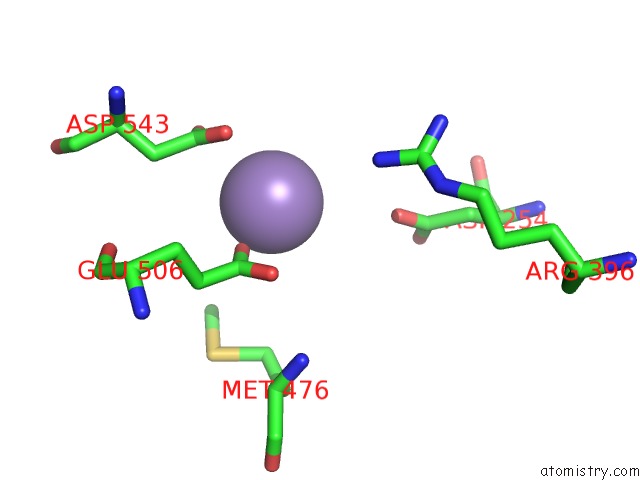
Mono view
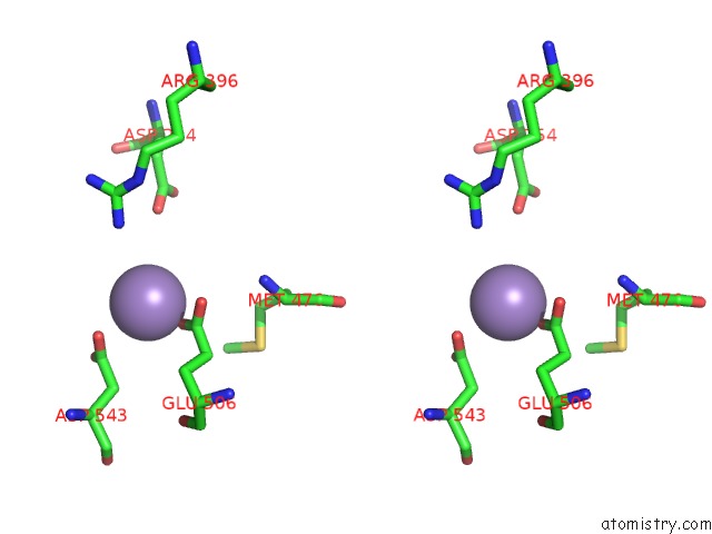
Stereo pair view
Mono view
Stereo pair view
A full contact list of Manganese with other atoms in the Mn binding
site number 1 of Crystal Structure of Mn(2+)-Bound Phosphoenolpyruvate Carboxylase within 5.0Å range:
|
Reference:
H.Matsumura,
M.Terada,
S.Shirakata,
T.Inoue,
T.Yoshinaga,
K.Izui,
Y.Kai.
Plausible Phosphoenolpyruvate Binding Site Revealed By 2.6 A Structure of MN2+-Bound Phosphoenolpyruvate Carboxylase From Escherichia Coli Febs Lett. V. 458 93 1999.
ISSN: ISSN 0014-5793
PubMed: 10481043
DOI: 10.1016/S0014-5793(99)01103-5
Page generated: Sat Oct 5 12:10:52 2024
ISSN: ISSN 0014-5793
PubMed: 10481043
DOI: 10.1016/S0014-5793(99)01103-5
Last articles
Mg in 3AQLMg in 3AQB
Mg in 3AOI
Mg in 3AOH
Mg in 3AQ4
Mg in 3ALO
Mg in 3AO2
Mg in 3AM1
Mg in 3AK8
Mg in 3ALN