Manganese »
PDB 1pj4-1r1o »
1pkn »
Manganese in PDB 1pkn: Structure of Rabbit Muscle Pyruvate Kinase Complexed with MN2+, K+, and Pyruvate
Enzymatic activity of Structure of Rabbit Muscle Pyruvate Kinase Complexed with MN2+, K+, and Pyruvate
All present enzymatic activity of Structure of Rabbit Muscle Pyruvate Kinase Complexed with MN2+, K+, and Pyruvate:
2.7.1.40;
2.7.1.40;
Protein crystallography data
The structure of Structure of Rabbit Muscle Pyruvate Kinase Complexed with MN2+, K+, and Pyruvate, PDB code: 1pkn
was solved by
T.M.Larsen,
L.T.Laughlin,
H.M.Holden,
I.Rayment,
G.H.Reed,
with X-Ray Crystallography technique. A brief refinement statistics is given in the table below:
| Resolution Low / High (Å) | 30.00 / 2.90 |
| Space group | P 1 |
| Cell size a, b, c (Å), α, β, γ (°) | 83.600, 109.900, 146.800, 94.90, 93.60, 112.30 |
| R / Rfree (%) | n/a / n/a |
Other elements in 1pkn:
The structure of Structure of Rabbit Muscle Pyruvate Kinase Complexed with MN2+, K+, and Pyruvate also contains other interesting chemical elements:
| Potassium | (K) | 1 atom |
Manganese Binding Sites:
The binding sites of Manganese atom in the Structure of Rabbit Muscle Pyruvate Kinase Complexed with MN2+, K+, and Pyruvate
(pdb code 1pkn). This binding sites where shown within
5.0 Angstroms radius around Manganese atom.
In total only one binding site of Manganese was determined in the Structure of Rabbit Muscle Pyruvate Kinase Complexed with MN2+, K+, and Pyruvate, PDB code: 1pkn:
In total only one binding site of Manganese was determined in the Structure of Rabbit Muscle Pyruvate Kinase Complexed with MN2+, K+, and Pyruvate, PDB code: 1pkn:
Manganese binding site 1 out of 1 in 1pkn
Go back to
Manganese binding site 1 out
of 1 in the Structure of Rabbit Muscle Pyruvate Kinase Complexed with MN2+, K+, and Pyruvate
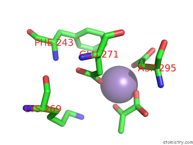
Mono view
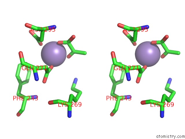
Stereo pair view
Mono view
Stereo pair view
A full contact list of Manganese with other atoms in the Mn binding
site number 1 of Structure of Rabbit Muscle Pyruvate Kinase Complexed with MN2+, K+, and Pyruvate within 5.0Å range:
|
Reference:
T.M.Larsen,
L.T.Laughlin,
H.M.Holden,
I.Rayment,
G.H.Reed.
Structure of Rabbit Muscle Pyruvate Kinase Complexed with MN2+, K+, and Pyruvate. Biochemistry V. 33 6301 1994.
ISSN: ISSN 0006-2960
PubMed: 8193145
DOI: 10.1021/BI00186A033
Page generated: Sat Oct 5 12:07:35 2024
ISSN: ISSN 0006-2960
PubMed: 8193145
DOI: 10.1021/BI00186A033
Last articles
Mg in 2UUBMg in 2UUC
Mg in 2UX5
Mg in 2UX4
Mg in 2UX3
Mg in 2UWW
Mg in 2UWV
Mg in 2UWU
Mg in 2UWT
Mg in 2UUA