Manganese »
PDB 1o9i-1pj3 »
1phk »
Manganese in PDB 1phk: Two Structures of the Catalytic Domain of Phosphorylase, Kinase: An Active Protein Kinase Complexed with Nucleotide, Substrate-Analogue and Product
Enzymatic activity of Two Structures of the Catalytic Domain of Phosphorylase, Kinase: An Active Protein Kinase Complexed with Nucleotide, Substrate-Analogue and Product
All present enzymatic activity of Two Structures of the Catalytic Domain of Phosphorylase, Kinase: An Active Protein Kinase Complexed with Nucleotide, Substrate-Analogue and Product:
2.7.1.38;
2.7.1.38;
Protein crystallography data
The structure of Two Structures of the Catalytic Domain of Phosphorylase, Kinase: An Active Protein Kinase Complexed with Nucleotide, Substrate-Analogue and Product, PDB code: 1phk
was solved by
D.J.Owen,
M.E.M.Noble,
E.F.Garman,
A.C.Papageorgiou,
L.N.Johnson,
with X-Ray Crystallography technique. A brief refinement statistics is given in the table below:
| Resolution Low / High (Å) | 6.00 / 2.20 |
| Space group | P 21 21 21 |
| Cell size a, b, c (Å), α, β, γ (°) | 47.600, 67.400, 110.800, 90.00, 90.00, 90.00 |
| R / Rfree (%) | 21 / 28.8 |
Manganese Binding Sites:
The binding sites of Manganese atom in the Two Structures of the Catalytic Domain of Phosphorylase, Kinase: An Active Protein Kinase Complexed with Nucleotide, Substrate-Analogue and Product
(pdb code 1phk). This binding sites where shown within
5.0 Angstroms radius around Manganese atom.
In total 2 binding sites of Manganese where determined in the Two Structures of the Catalytic Domain of Phosphorylase, Kinase: An Active Protein Kinase Complexed with Nucleotide, Substrate-Analogue and Product, PDB code: 1phk:
Jump to Manganese binding site number: 1; 2;
In total 2 binding sites of Manganese where determined in the Two Structures of the Catalytic Domain of Phosphorylase, Kinase: An Active Protein Kinase Complexed with Nucleotide, Substrate-Analogue and Product, PDB code: 1phk:
Jump to Manganese binding site number: 1; 2;
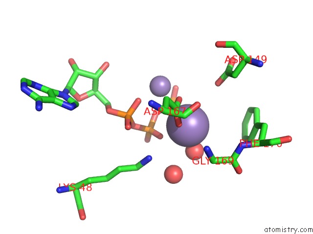
Manganese binding site 1 out of 2 in 1phk
Go back to
Manganese binding site 1 out
of 2 in the Two Structures of the Catalytic Domain of Phosphorylase, Kinase: An Active Protein Kinase Complexed with Nucleotide, Substrate-Analogue and Product
Mono view
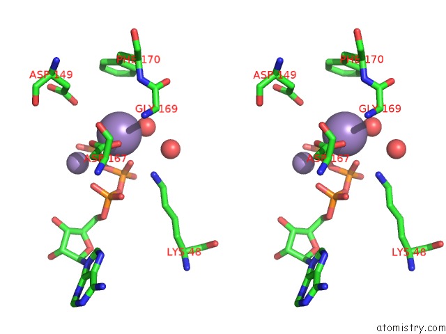
Stereo pair view
Mono view
Stereo pair view
A full contact list of Manganese with other atoms in the Mn binding
site number 1 of Two Structures of the Catalytic Domain of Phosphorylase, Kinase: An Active Protein Kinase Complexed with Nucleotide, Substrate-Analogue and Product within 5.0Å range:
|
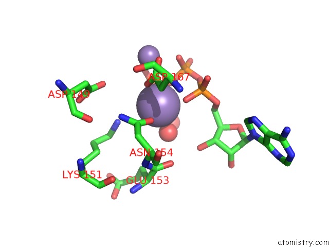
Manganese binding site 2 out of 2 in 1phk
Go back to
Manganese binding site 2 out
of 2 in the Two Structures of the Catalytic Domain of Phosphorylase, Kinase: An Active Protein Kinase Complexed with Nucleotide, Substrate-Analogue and Product
Mono view
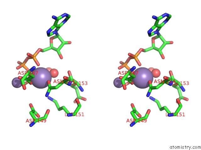
Stereo pair view
Mono view
Stereo pair view
A full contact list of Manganese with other atoms in the Mn binding
site number 2 of Two Structures of the Catalytic Domain of Phosphorylase, Kinase: An Active Protein Kinase Complexed with Nucleotide, Substrate-Analogue and Product within 5.0Å range:
|
Reference:
D.J.Owen,
M.E.Noble,
E.F.Garman,
A.C.Papageorgiou,
L.N.Johnson.
Two Structures of the Catalytic Domain of Phosphorylase Kinase: An Active Protein Kinase Complexed with Substrate Analogue and Product. Structure V. 3 467 1995.
ISSN: ISSN 0969-2126
PubMed: 7663944
DOI: 10.1016/S0969-2126(01)00180-0
Page generated: Sat Oct 5 12:05:16 2024
ISSN: ISSN 0969-2126
PubMed: 7663944
DOI: 10.1016/S0969-2126(01)00180-0
Last articles
K in 3HHEK in 3HOG
K in 3HJW
K in 3HFW
K in 3HC1
K in 3GVX
K in 3H7C
K in 3GX3
K in 3H4K
K in 3GX5