Manganese »
PDB 1n0n-1o99 »
1n1p »
Manganese in PDB 1n1p: Atomic Resolution Structure of Cholesterol Oxidase @ pH 7.4 (Streptomyces Sp. Sa-Coo)
Enzymatic activity of Atomic Resolution Structure of Cholesterol Oxidase @ pH 7.4 (Streptomyces Sp. Sa-Coo)
All present enzymatic activity of Atomic Resolution Structure of Cholesterol Oxidase @ pH 7.4 (Streptomyces Sp. Sa-Coo):
1.1.3.6;
1.1.3.6;
Protein crystallography data
The structure of Atomic Resolution Structure of Cholesterol Oxidase @ pH 7.4 (Streptomyces Sp. Sa-Coo), PDB code: 1n1p
was solved by
A.Vrielink,
P.I.Lario,
with X-Ray Crystallography technique. A brief refinement statistics is given in the table below:
| Resolution Low / High (Å) | 49.00 / 0.95 |
| Space group | P 1 21 1 |
| Cell size a, b, c (Å), α, β, γ (°) | 51.273, 72.964, 63.036, 90.00, 105.18, 90.00 |
| R / Rfree (%) | 9.7 / 11.9 |
Manganese Binding Sites:
The binding sites of Manganese atom in the Atomic Resolution Structure of Cholesterol Oxidase @ pH 7.4 (Streptomyces Sp. Sa-Coo)
(pdb code 1n1p). This binding sites where shown within
5.0 Angstroms radius around Manganese atom.
In total 3 binding sites of Manganese where determined in the Atomic Resolution Structure of Cholesterol Oxidase @ pH 7.4 (Streptomyces Sp. Sa-Coo), PDB code: 1n1p:
Jump to Manganese binding site number: 1; 2; 3;
In total 3 binding sites of Manganese where determined in the Atomic Resolution Structure of Cholesterol Oxidase @ pH 7.4 (Streptomyces Sp. Sa-Coo), PDB code: 1n1p:
Jump to Manganese binding site number: 1; 2; 3;
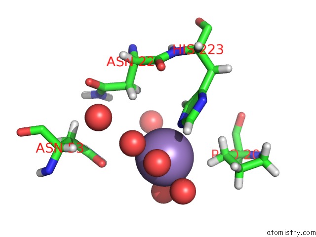
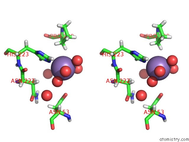
Manganese binding site 1 out of 3 in 1n1p
Go back to
Manganese binding site 1 out
of 3 in the Atomic Resolution Structure of Cholesterol Oxidase @ pH 7.4 (Streptomyces Sp. Sa-Coo)
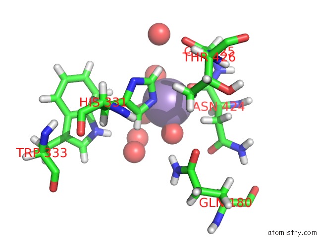
Mono view
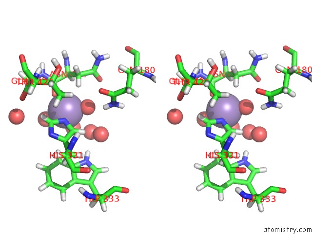
Stereo pair view
Mono view
Stereo pair view
A full contact list of Manganese with other atoms in the Mn binding
site number 1 of Atomic Resolution Structure of Cholesterol Oxidase @ pH 7.4 (Streptomyces Sp. Sa-Coo) within 5.0Å range:
|
Manganese binding site 2 out of 3 in 1n1p
Go back to
Manganese binding site 2 out
of 3 in the Atomic Resolution Structure of Cholesterol Oxidase @ pH 7.4 (Streptomyces Sp. Sa-Coo)
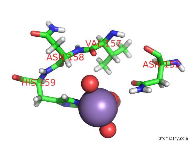
Mono view
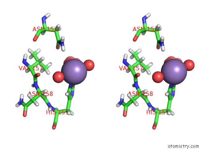
Stereo pair view
Mono view
Stereo pair view
A full contact list of Manganese with other atoms in the Mn binding
site number 2 of Atomic Resolution Structure of Cholesterol Oxidase @ pH 7.4 (Streptomyces Sp. Sa-Coo) within 5.0Å range:
|
Manganese binding site 3 out of 3 in 1n1p
Go back to
Manganese binding site 3 out
of 3 in the Atomic Resolution Structure of Cholesterol Oxidase @ pH 7.4 (Streptomyces Sp. Sa-Coo)
Mono view
Stereo pair view
Mono view
Stereo pair view
A full contact list of Manganese with other atoms in the Mn binding
site number 3 of Atomic Resolution Structure of Cholesterol Oxidase @ pH 7.4 (Streptomyces Sp. Sa-Coo) within 5.0Å range:
|
Reference:
P.I.Lario,
A.Vrielink.
Atomic Resolution Density Maps Reveal Secondary Structure Dependent Differences in Electronic Distribution J.Am.Chem.Soc. V. 125 12787 2003.
ISSN: ISSN 0002-7863
PubMed: 14558826
DOI: 10.1021/JA0289954
Page generated: Sat Oct 5 11:48:43 2024
ISSN: ISSN 0002-7863
PubMed: 14558826
DOI: 10.1021/JA0289954
Last articles
Mg in 3U0OMg in 3U08
Mg in 3U06
Mg in 3TY9
Mg in 3TZF
Mg in 3U05
Mg in 3TYZ
Mg in 3TZ5
Mg in 3TZ4
Mg in 3TXA