Manganese »
PDB 1lu1-1n0j »
1lzi »
Manganese in PDB 1lzi: Glycosyltransferase A + Udp + H Antigen Acceptor
Enzymatic activity of Glycosyltransferase A + Udp + H Antigen Acceptor
All present enzymatic activity of Glycosyltransferase A + Udp + H Antigen Acceptor:
2.4.1.40;
2.4.1.40;
Protein crystallography data
The structure of Glycosyltransferase A + Udp + H Antigen Acceptor, PDB code: 1lzi
was solved by
S.I.Patenaude,
N.O.L.Seto,
S.N.Borisova,
A.Szpacenko,
S.L.Marcus,
M.M.Palcic,
S.V.Evans,
with X-Ray Crystallography technique. A brief refinement statistics is given in the table below:
| Resolution Low / High (Å) | 20.00 / 1.35 |
| Space group | C 2 2 21 |
| Cell size a, b, c (Å), α, β, γ (°) | 52.810, 149.580, 79.970, 90.00, 90.00, 90.00 |
| R / Rfree (%) | 17.9 / 20 |
Other elements in 1lzi:
The structure of Glycosyltransferase A + Udp + H Antigen Acceptor also contains other interesting chemical elements:
| Mercury | (Hg) | 5 atoms |
Manganese Binding Sites:
The binding sites of Manganese atom in the Glycosyltransferase A + Udp + H Antigen Acceptor
(pdb code 1lzi). This binding sites where shown within
5.0 Angstroms radius around Manganese atom.
In total only one binding site of Manganese was determined in the Glycosyltransferase A + Udp + H Antigen Acceptor, PDB code: 1lzi:
In total only one binding site of Manganese was determined in the Glycosyltransferase A + Udp + H Antigen Acceptor, PDB code: 1lzi:
Manganese binding site 1 out of 1 in 1lzi
Go back to
Manganese binding site 1 out
of 1 in the Glycosyltransferase A + Udp + H Antigen Acceptor
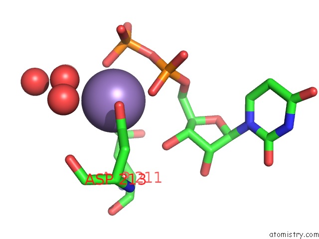
Mono view
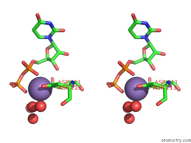
Stereo pair view
Mono view
Stereo pair view
A full contact list of Manganese with other atoms in the Mn binding
site number 1 of Glycosyltransferase A + Udp + H Antigen Acceptor within 5.0Å range:
|
Reference:
S.I.Patenaude,
N.O.Seto,
S.N.Borisova,
A.Szpacenko,
S.L.Marcus,
M.M.Palcic,
S.V.Evans.
The Structural Basis For Specificity in Human Abo(H) Blood Group Biosynthesis. Nat.Struct.Biol. V. 9 685 2002.
ISSN: ISSN 1072-8368
PubMed: 12198488
DOI: 10.1038/NSB832
Page generated: Sat Oct 5 11:37:08 2024
ISSN: ISSN 1072-8368
PubMed: 12198488
DOI: 10.1038/NSB832
Last articles
K in 4UA6K in 4UD8
K in 4U76
K in 4U75
K in 4U73
K in 4U71
K in 4U70
K in 4U6Z
K in 4TS0
K in 4U6W