Manganese »
PDB 1khe-1lte »
1lqk »
Manganese in PDB 1lqk: High Resolution Structure of Fosfomycin Resistance Protein A (Fosa)
Enzymatic activity of High Resolution Structure of Fosfomycin Resistance Protein A (Fosa)
All present enzymatic activity of High Resolution Structure of Fosfomycin Resistance Protein A (Fosa):
2.5.1.18;
2.5.1.18;
Protein crystallography data
The structure of High Resolution Structure of Fosfomycin Resistance Protein A (Fosa), PDB code: 1lqk
was solved by
C.L.Rife,
R.E.Pharris,
M.E.Newcomer,
R.N.Armstrong,
with X-Ray Crystallography technique. A brief refinement statistics is given in the table below:
| Resolution Low / High (Å) | 30.00 / 1.35 |
| Space group | P 21 21 21 |
| Cell size a, b, c (Å), α, β, γ (°) | 54.788, 66.967, 77.000, 90.00, 90.00, 90.00 |
| R / Rfree (%) | 14.3 / 18.5 |
Other elements in 1lqk:
The structure of High Resolution Structure of Fosfomycin Resistance Protein A (Fosa) also contains other interesting chemical elements:
| Potassium | (K) | 2 atoms |
Manganese Binding Sites:
The binding sites of Manganese atom in the High Resolution Structure of Fosfomycin Resistance Protein A (Fosa)
(pdb code 1lqk). This binding sites where shown within
5.0 Angstroms radius around Manganese atom.
In total 2 binding sites of Manganese where determined in the High Resolution Structure of Fosfomycin Resistance Protein A (Fosa), PDB code: 1lqk:
Jump to Manganese binding site number: 1; 2;
In total 2 binding sites of Manganese where determined in the High Resolution Structure of Fosfomycin Resistance Protein A (Fosa), PDB code: 1lqk:
Jump to Manganese binding site number: 1; 2;
Manganese binding site 1 out of 2 in 1lqk
Go back to
Manganese binding site 1 out
of 2 in the High Resolution Structure of Fosfomycin Resistance Protein A (Fosa)
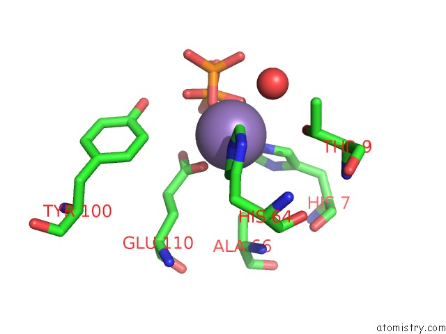
Mono view
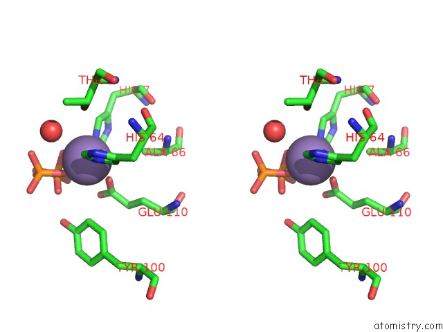
Stereo pair view
Mono view
Stereo pair view
A full contact list of Manganese with other atoms in the Mn binding
site number 1 of High Resolution Structure of Fosfomycin Resistance Protein A (Fosa) within 5.0Å range:
|
Manganese binding site 2 out of 2 in 1lqk
Go back to
Manganese binding site 2 out
of 2 in the High Resolution Structure of Fosfomycin Resistance Protein A (Fosa)
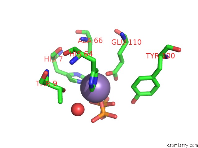
Mono view
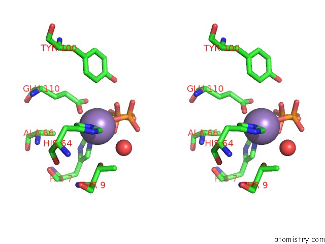
Stereo pair view
Mono view
Stereo pair view
A full contact list of Manganese with other atoms in the Mn binding
site number 2 of High Resolution Structure of Fosfomycin Resistance Protein A (Fosa) within 5.0Å range:
|
Reference:
C.L.Rife,
R.E.Pharris,
M.E.Newcomer,
R.N.Armstrong.
Crystal Structure of A Genomically Encoded Fosfomycin Resistance Protein (Fosa) at 1.19 A Resolution By Mad Phasing Off the L-III Edge of Tl(+) J.Am.Chem.Soc. V. 124 11001 2002.
ISSN: ISSN 0002-7863
PubMed: 12224946
DOI: 10.1021/JA026879V
Page generated: Sat Oct 5 11:33:19 2024
ISSN: ISSN 0002-7863
PubMed: 12224946
DOI: 10.1021/JA026879V
Last articles
I in 4F4BI in 4E27
I in 4EUU
I in 4EUT
I in 4EBK
I in 4E9O
I in 4DZL
I in 4E91
I in 4DZN
I in 4DZM