Manganese »
PDB 1khe-1lte »
1lnc »
Manganese in PDB 1lnc: A Structural Analysis of Metal Substitutions in Thermolysin
Enzymatic activity of A Structural Analysis of Metal Substitutions in Thermolysin
All present enzymatic activity of A Structural Analysis of Metal Substitutions in Thermolysin:
3.4.24.27;
3.4.24.27;
Protein crystallography data
The structure of A Structural Analysis of Metal Substitutions in Thermolysin, PDB code: 1lnc
was solved by
D.R.Holland,
A.C.Hausrath,
D.Juers,
B.W.Matthews,
with X-Ray Crystallography technique. A brief refinement statistics is given in the table below:
| Resolution Low / High (Å) | 20.00 / 1.80 |
| Space group | P 61 2 2 |
| Cell size a, b, c (Å), α, β, γ (°) | 93.900, 93.900, 131.200, 90.00, 90.00, 120.00 |
| R / Rfree (%) | n/a / n/a |
Other elements in 1lnc:
The structure of A Structural Analysis of Metal Substitutions in Thermolysin also contains other interesting chemical elements:
| Calcium | (Ca) | 2 atoms |
Manganese Binding Sites:
The binding sites of Manganese atom in the A Structural Analysis of Metal Substitutions in Thermolysin
(pdb code 1lnc). This binding sites where shown within
5.0 Angstroms radius around Manganese atom.
In total 3 binding sites of Manganese where determined in the A Structural Analysis of Metal Substitutions in Thermolysin, PDB code: 1lnc:
Jump to Manganese binding site number: 1; 2; 3;
In total 3 binding sites of Manganese where determined in the A Structural Analysis of Metal Substitutions in Thermolysin, PDB code: 1lnc:
Jump to Manganese binding site number: 1; 2; 3;
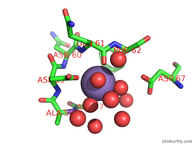
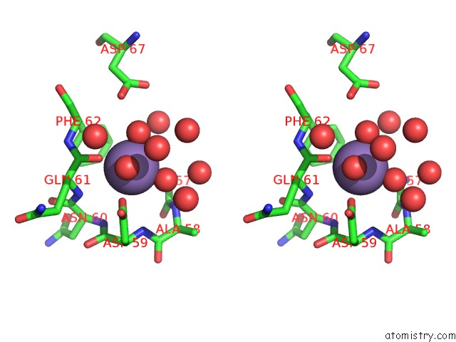
Manganese binding site 1 out of 3 in 1lnc
Go back to
Manganese binding site 1 out
of 3 in the A Structural Analysis of Metal Substitutions in Thermolysin
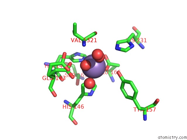
Mono view
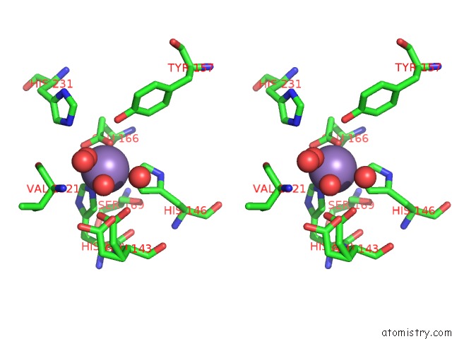
Stereo pair view
Mono view
Stereo pair view
A full contact list of Manganese with other atoms in the Mn binding
site number 1 of A Structural Analysis of Metal Substitutions in Thermolysin within 5.0Å range:
|
Manganese binding site 2 out of 3 in 1lnc
Go back to
Manganese binding site 2 out
of 3 in the A Structural Analysis of Metal Substitutions in Thermolysin
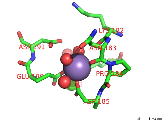
Mono view
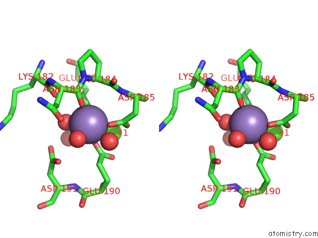
Stereo pair view
Mono view
Stereo pair view
A full contact list of Manganese with other atoms in the Mn binding
site number 2 of A Structural Analysis of Metal Substitutions in Thermolysin within 5.0Å range:
|
Manganese binding site 3 out of 3 in 1lnc
Go back to
Manganese binding site 3 out
of 3 in the A Structural Analysis of Metal Substitutions in Thermolysin
Mono view
Stereo pair view
Mono view
Stereo pair view
A full contact list of Manganese with other atoms in the Mn binding
site number 3 of A Structural Analysis of Metal Substitutions in Thermolysin within 5.0Å range:
|
Reference:
D.R.Holland,
A.C.Hausrath,
D.Juers,
B.W.Matthews.
Structural Analysis of Zinc Substitutions in the Active Site of Thermolysin. Protein Sci. V. 4 1955 1995.
ISSN: ISSN 0961-8368
PubMed: 8535232
Page generated: Sat Oct 5 11:30:05 2024
ISSN: ISSN 0961-8368
PubMed: 8535232
Last articles
K in 1G8MK in 1GA9
K in 1FPI
K in 1G75
K in 1F3X
K in 1FT7
K in 1FQE
K in 1F3W
K in 1FP7
K in 1FL1