Manganese »
PDB 1khe-1lte »
1ll2 »
Manganese in PDB 1ll2: Crystal Structure of Rabbit Muscle Glycogenin Complexed with Udp- Glucose and Manganese
Enzymatic activity of Crystal Structure of Rabbit Muscle Glycogenin Complexed with Udp- Glucose and Manganese
All present enzymatic activity of Crystal Structure of Rabbit Muscle Glycogenin Complexed with Udp- Glucose and Manganese:
2.4.1.186;
2.4.1.186;
Protein crystallography data
The structure of Crystal Structure of Rabbit Muscle Glycogenin Complexed with Udp- Glucose and Manganese, PDB code: 1ll2
was solved by
B.J.Gibbons,
P.J.Roach,
T.D.Hurley,
with X-Ray Crystallography technique. A brief refinement statistics is given in the table below:
| Resolution Low / High (Å) | 30.00 / 1.90 |
| Space group | I 2 2 2 |
| Cell size a, b, c (Å), α, β, γ (°) | 57.887, 106.875, 122.253, 90.00, 90.00, 90.00 |
| R / Rfree (%) | 19.4 / 22.7 |
Manganese Binding Sites:
The binding sites of Manganese atom in the Crystal Structure of Rabbit Muscle Glycogenin Complexed with Udp- Glucose and Manganese
(pdb code 1ll2). This binding sites where shown within
5.0 Angstroms radius around Manganese atom.
In total only one binding site of Manganese was determined in the Crystal Structure of Rabbit Muscle Glycogenin Complexed with Udp- Glucose and Manganese, PDB code: 1ll2:
In total only one binding site of Manganese was determined in the Crystal Structure of Rabbit Muscle Glycogenin Complexed with Udp- Glucose and Manganese, PDB code: 1ll2:
Manganese binding site 1 out of 1 in 1ll2
Go back to
Manganese binding site 1 out
of 1 in the Crystal Structure of Rabbit Muscle Glycogenin Complexed with Udp- Glucose and Manganese
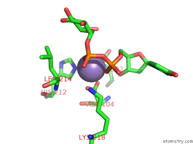
Mono view
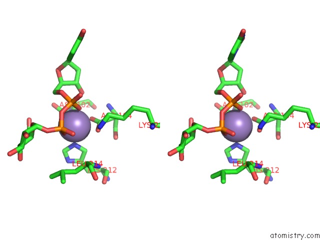
Stereo pair view
Mono view
Stereo pair view
A full contact list of Manganese with other atoms in the Mn binding
site number 1 of Crystal Structure of Rabbit Muscle Glycogenin Complexed with Udp- Glucose and Manganese within 5.0Å range:
|
Reference:
B.J.Gibbons,
P.J.Roach,
T.D.Hurley.
Crystal Structure of the Autocatalytic Initiator of Glycogen Biosynthesis, Glycogenin. J.Mol.Biol. V. 319 463 2002.
ISSN: ISSN 0022-2836
PubMed: 12051921
DOI: 10.1016/S0022-2836(02)00305-4
Page generated: Sat Oct 5 11:29:57 2024
ISSN: ISSN 0022-2836
PubMed: 12051921
DOI: 10.1016/S0022-2836(02)00305-4
Last articles
K in 2VWJK in 2VXY
K in 2VQW
K in 2VQV
K in 2VQQ
K in 2VQO
K in 2VI5
K in 2VQM
K in 2VQJ
K in 2VPL