Manganese »
PDB 1khe-1lte »
1ksi »
Manganese in PDB 1ksi: Crystal Structure of A Eukaryotic (Pea Seedling) Copper-Containing Amine Oxidase at 2.2A Resolution
Enzymatic activity of Crystal Structure of A Eukaryotic (Pea Seedling) Copper-Containing Amine Oxidase at 2.2A Resolution
All present enzymatic activity of Crystal Structure of A Eukaryotic (Pea Seedling) Copper-Containing Amine Oxidase at 2.2A Resolution:
1.4.3.6;
1.4.3.6;
Protein crystallography data
The structure of Crystal Structure of A Eukaryotic (Pea Seedling) Copper-Containing Amine Oxidase at 2.2A Resolution, PDB code: 1ksi
was solved by
M.C.J.Wilce,
V.Kumar,
H.C.Freeman,
J.M.Guss,
with X-Ray Crystallography technique. A brief refinement statistics is given in the table below:
| Resolution Low / High (Å) | 7.00 / 2.20 |
| Space group | P 21 21 21 |
| Cell size a, b, c (Å), α, β, γ (°) | 85.370, 114.640, 199.940, 90.00, 90.00, 90.00 |
| R / Rfree (%) | n/a / n/a |
Other elements in 1ksi:
The structure of Crystal Structure of A Eukaryotic (Pea Seedling) Copper-Containing Amine Oxidase at 2.2A Resolution also contains other interesting chemical elements:
| Copper | (Cu) | 2 atoms |
Manganese Binding Sites:
The binding sites of Manganese atom in the Crystal Structure of A Eukaryotic (Pea Seedling) Copper-Containing Amine Oxidase at 2.2A Resolution
(pdb code 1ksi). This binding sites where shown within
5.0 Angstroms radius around Manganese atom.
In total 2 binding sites of Manganese where determined in the Crystal Structure of A Eukaryotic (Pea Seedling) Copper-Containing Amine Oxidase at 2.2A Resolution, PDB code: 1ksi:
Jump to Manganese binding site number: 1; 2;
In total 2 binding sites of Manganese where determined in the Crystal Structure of A Eukaryotic (Pea Seedling) Copper-Containing Amine Oxidase at 2.2A Resolution, PDB code: 1ksi:
Jump to Manganese binding site number: 1; 2;
Manganese binding site 1 out of 2 in 1ksi
Go back to
Manganese binding site 1 out
of 2 in the Crystal Structure of A Eukaryotic (Pea Seedling) Copper-Containing Amine Oxidase at 2.2A Resolution
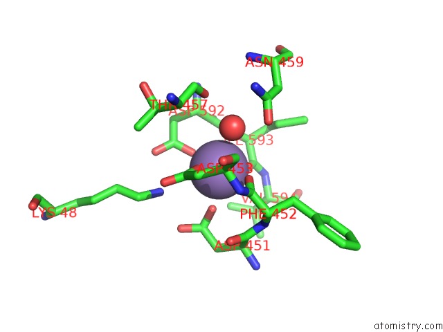
Mono view
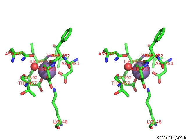
Stereo pair view
Mono view
Stereo pair view
A full contact list of Manganese with other atoms in the Mn binding
site number 1 of Crystal Structure of A Eukaryotic (Pea Seedling) Copper-Containing Amine Oxidase at 2.2A Resolution within 5.0Å range:
|
Manganese binding site 2 out of 2 in 1ksi
Go back to
Manganese binding site 2 out
of 2 in the Crystal Structure of A Eukaryotic (Pea Seedling) Copper-Containing Amine Oxidase at 2.2A Resolution
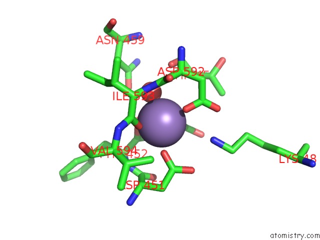
Mono view
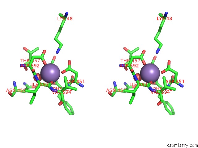
Stereo pair view
Mono view
Stereo pair view
A full contact list of Manganese with other atoms in the Mn binding
site number 2 of Crystal Structure of A Eukaryotic (Pea Seedling) Copper-Containing Amine Oxidase at 2.2A Resolution within 5.0Å range:
|
Reference:
V.Kumar,
D.M.Dooley,
H.C.Freeman,
J.M.Guss,
I.Harvey,
M.A.Mcguirl,
M.C.Wilce,
V.M.Zubak.
Crystal Structure of A Eukaryotic (Pea Seedling) Copper-Containing Amine Oxidase at 2.2 A Resolution. Structure V. 4 943 1996.
ISSN: ISSN 0969-2126
PubMed: 8805580
DOI: 10.1016/S0969-2126(96)00101-3
Page generated: Sat Oct 5 11:23:53 2024
ISSN: ISSN 0969-2126
PubMed: 8805580
DOI: 10.1016/S0969-2126(96)00101-3
Last articles
I in 2AK4I in 2ARL
I in 2ANX
I in 2AQW
I in 2AF6
I in 2ANV
I in 2A0N
I in 1XC6
I in 1ZVV
I in 1Z7J