Manganese »
PDB 1j54-1khb »
1kfl »
Manganese in PDB 1kfl: Crystal Structure of Phenylalanine-Regulated 3-Deoxy-D- Arabino-Heptulosonate-7-Phosphate Synthase (Dahp Synthase) From E.Coli Complexed with MN2+, Pep, and Phe
Enzymatic activity of Crystal Structure of Phenylalanine-Regulated 3-Deoxy-D- Arabino-Heptulosonate-7-Phosphate Synthase (Dahp Synthase) From E.Coli Complexed with MN2+, Pep, and Phe
All present enzymatic activity of Crystal Structure of Phenylalanine-Regulated 3-Deoxy-D- Arabino-Heptulosonate-7-Phosphate Synthase (Dahp Synthase) From E.Coli Complexed with MN2+, Pep, and Phe:
4.1.2.15;
4.1.2.15;
Protein crystallography data
The structure of Crystal Structure of Phenylalanine-Regulated 3-Deoxy-D- Arabino-Heptulosonate-7-Phosphate Synthase (Dahp Synthase) From E.Coli Complexed with MN2+, Pep, and Phe, PDB code: 1kfl
was solved by
I.A.Shumilin,
C.Zhao,
R.Bauerle,
R.H.Kretsinger,
with X-Ray Crystallography technique. A brief refinement statistics is given in the table below:
| Resolution Low / High (Å) | 20.00 / 2.80 |
| Space group | C 1 2 1 |
| Cell size a, b, c (Å), α, β, γ (°) | 290.097, 90.097, 155.798, 90.00, 120.77, 90.00 |
| R / Rfree (%) | 21.8 / 24.6 |
Manganese Binding Sites:
The binding sites of Manganese atom in the Crystal Structure of Phenylalanine-Regulated 3-Deoxy-D- Arabino-Heptulosonate-7-Phosphate Synthase (Dahp Synthase) From E.Coli Complexed with MN2+, Pep, and Phe
(pdb code 1kfl). This binding sites where shown within
5.0 Angstroms radius around Manganese atom.
In total 8 binding sites of Manganese where determined in the Crystal Structure of Phenylalanine-Regulated 3-Deoxy-D- Arabino-Heptulosonate-7-Phosphate Synthase (Dahp Synthase) From E.Coli Complexed with MN2+, Pep, and Phe, PDB code: 1kfl:
Jump to Manganese binding site number: 1; 2; 3; 4; 5; 6; 7; 8;
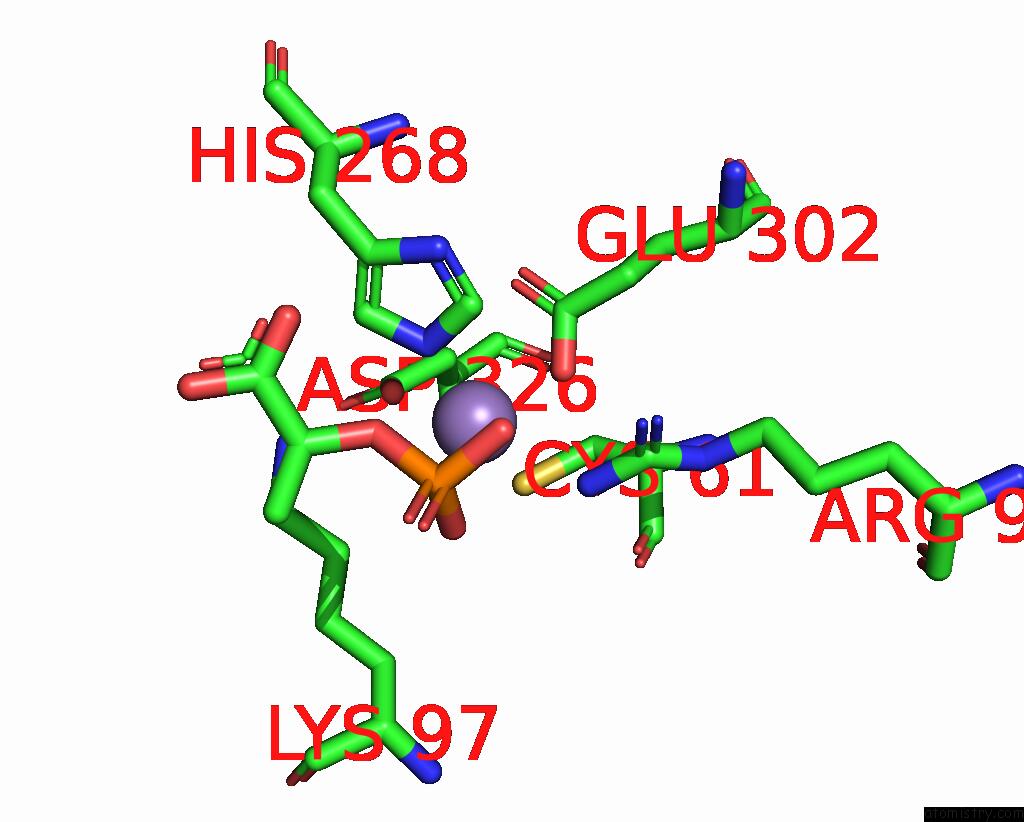
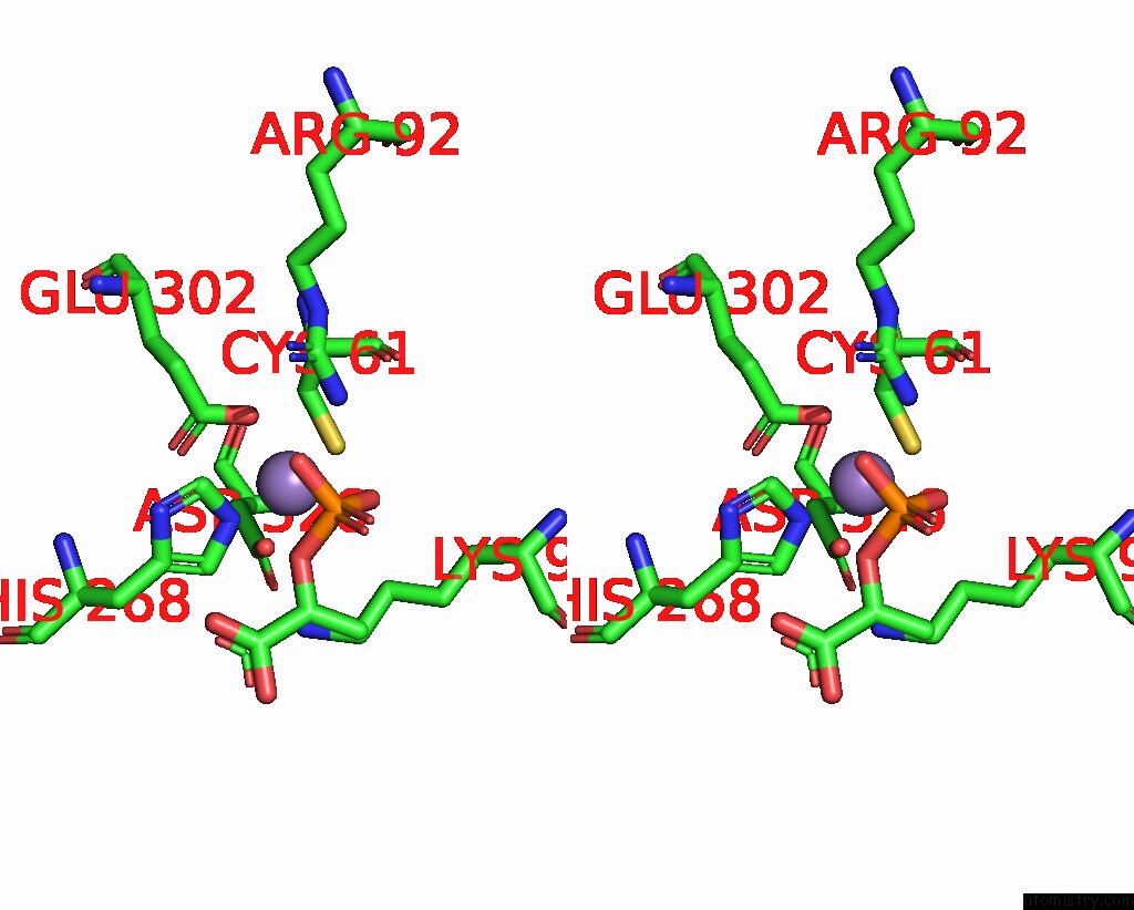
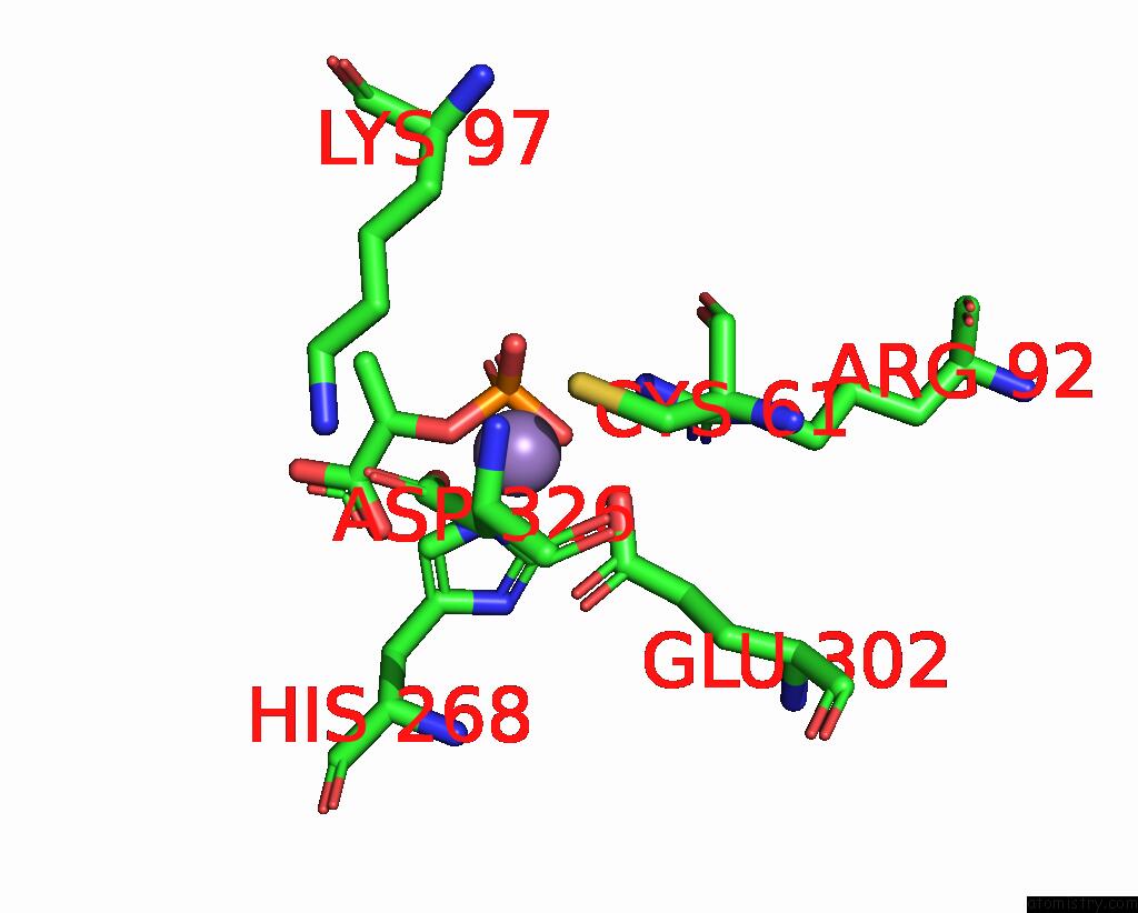
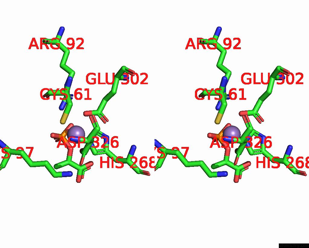
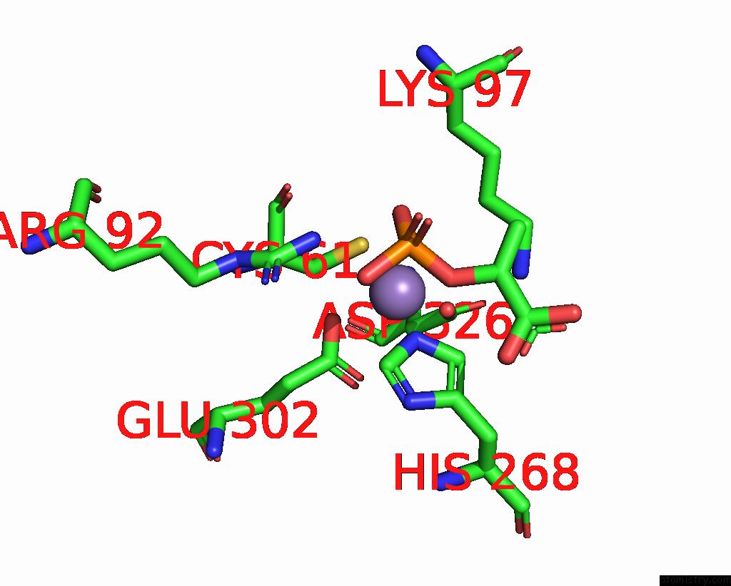
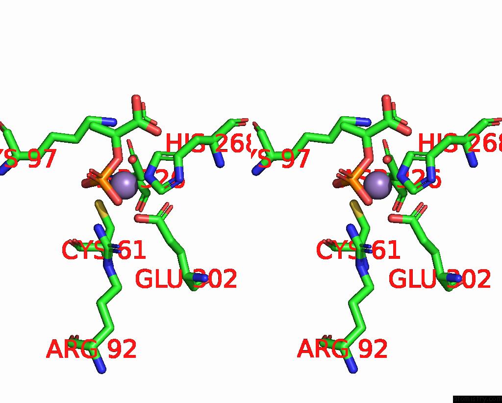
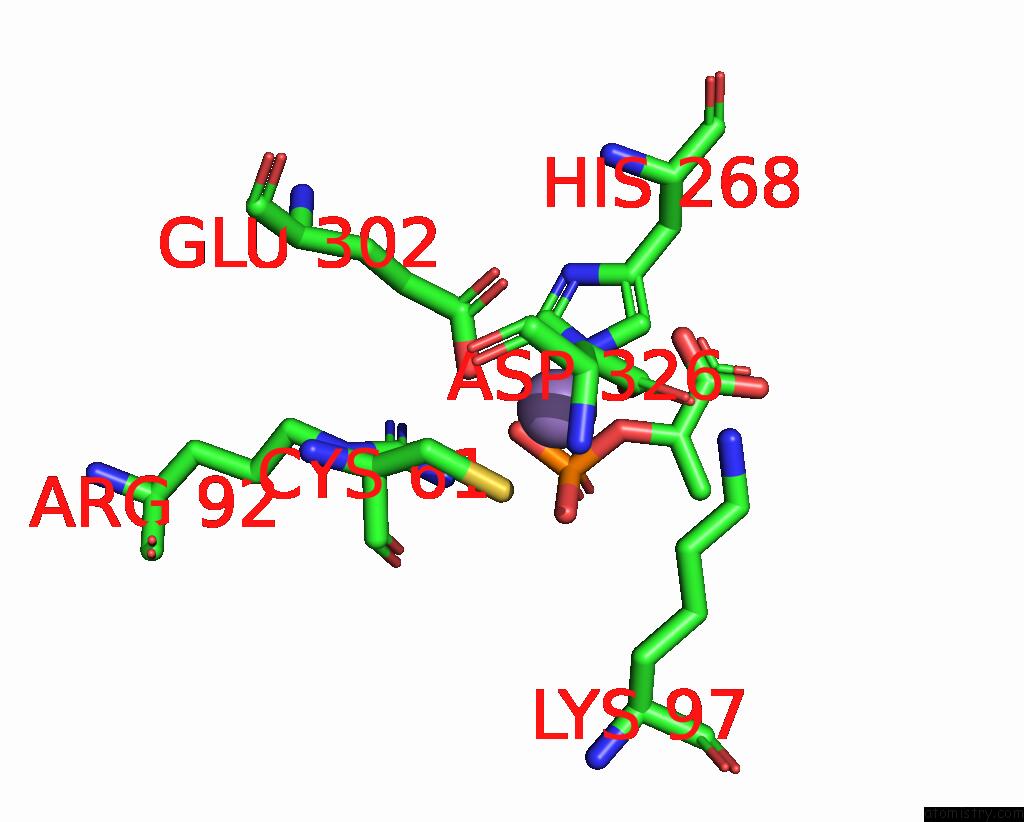
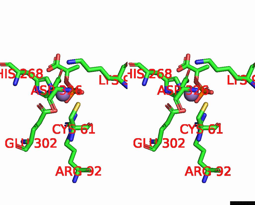
Manganese binding site 1 out of 8 in 1kfl
Go back to
Manganese binding site 1 out
of 8 in the Crystal Structure of Phenylalanine-Regulated 3-Deoxy-D- Arabino-Heptulosonate-7-Phosphate Synthase (Dahp Synthase) From E.Coli Complexed with MN2+, Pep, and Phe
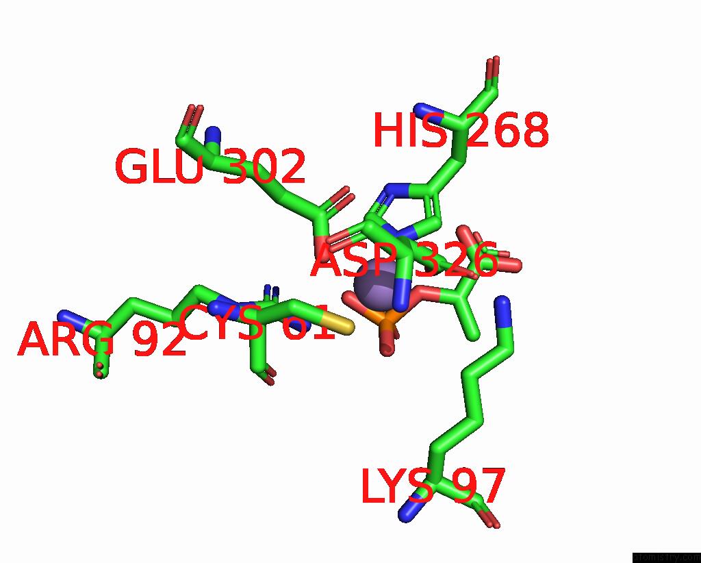
Mono view
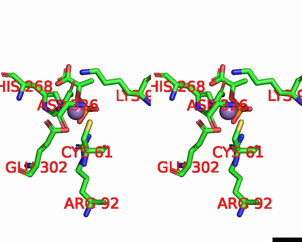
Stereo pair view
Mono view
Stereo pair view
A full contact list of Manganese with other atoms in the Mn binding
site number 1 of Crystal Structure of Phenylalanine-Regulated 3-Deoxy-D- Arabino-Heptulosonate-7-Phosphate Synthase (Dahp Synthase) From E.Coli Complexed with MN2+, Pep, and Phe within 5.0Å range:
|
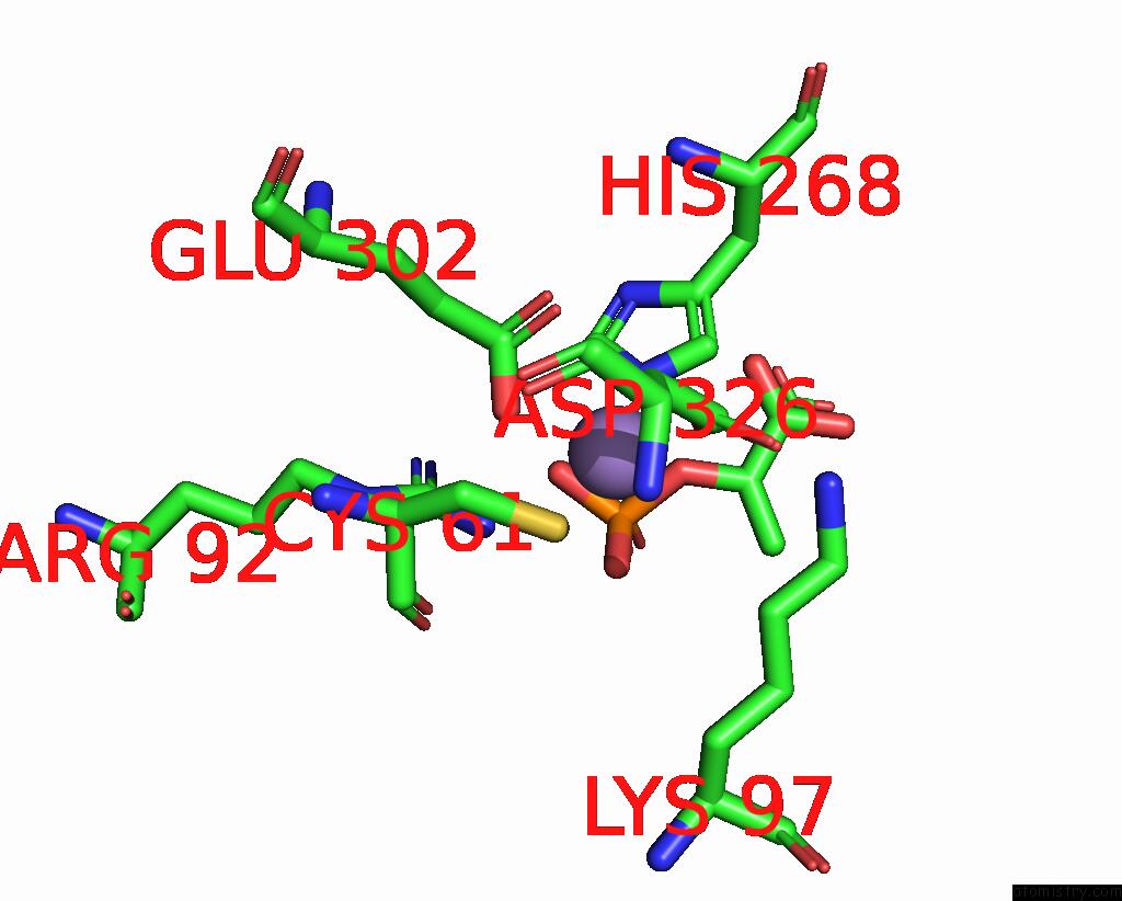
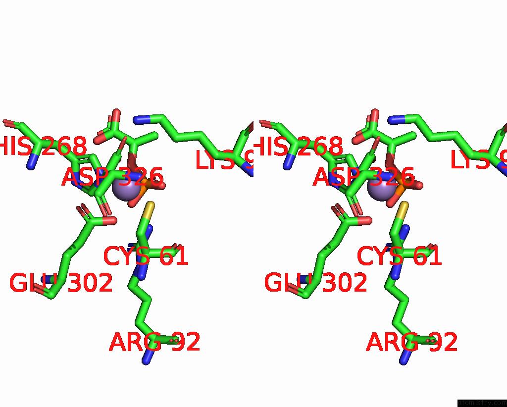
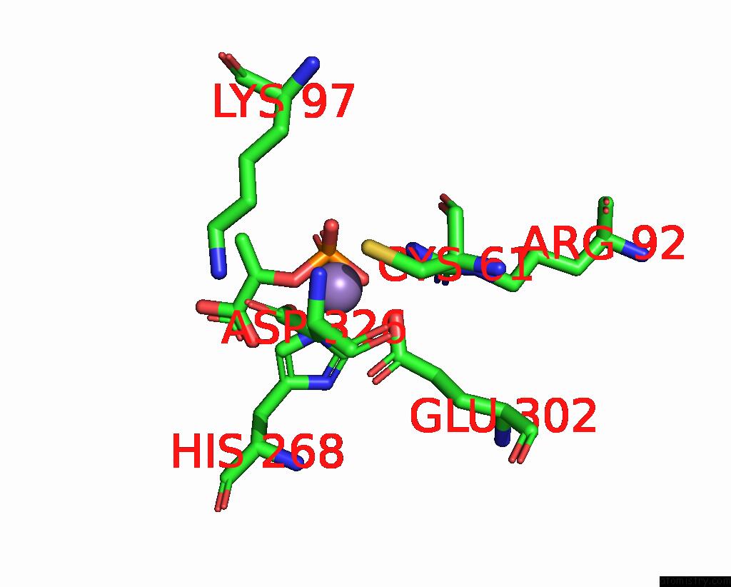
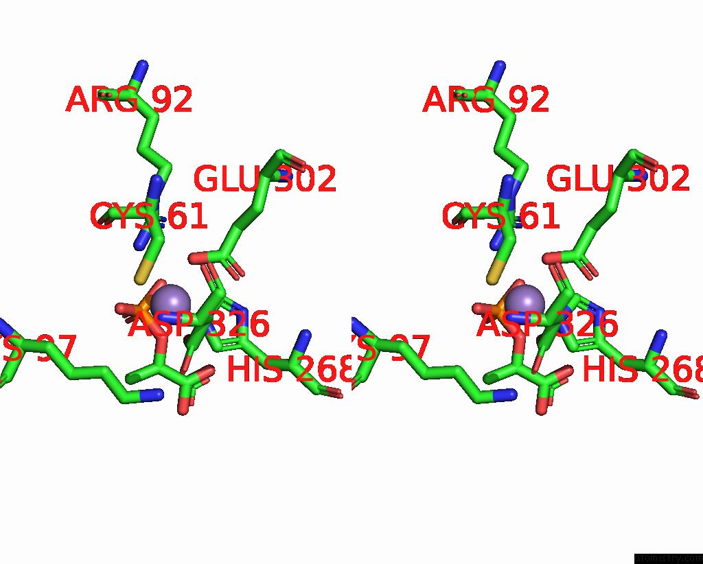
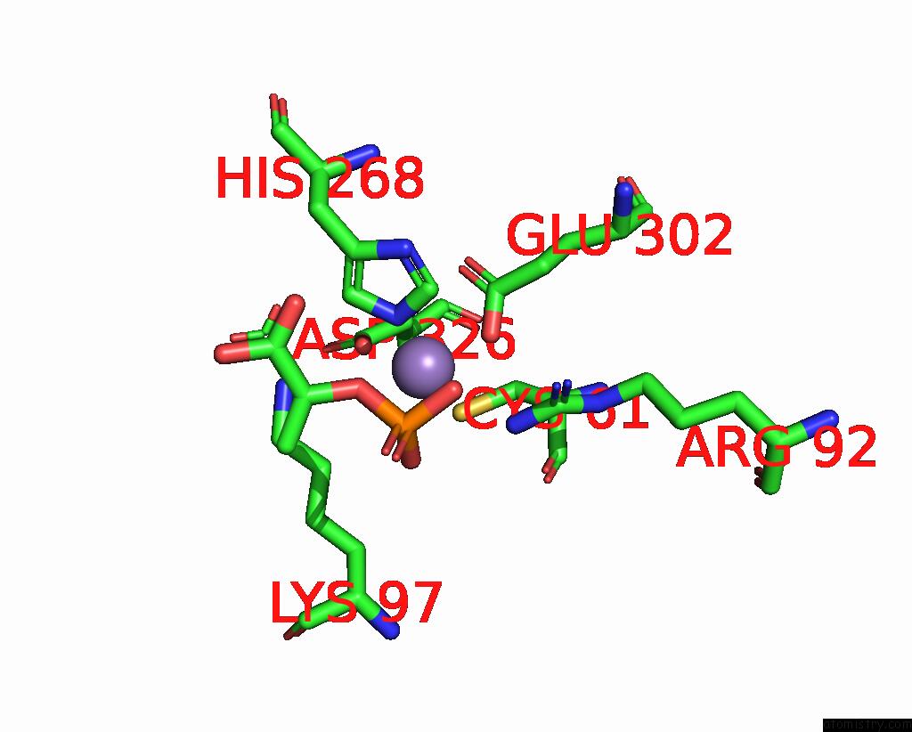
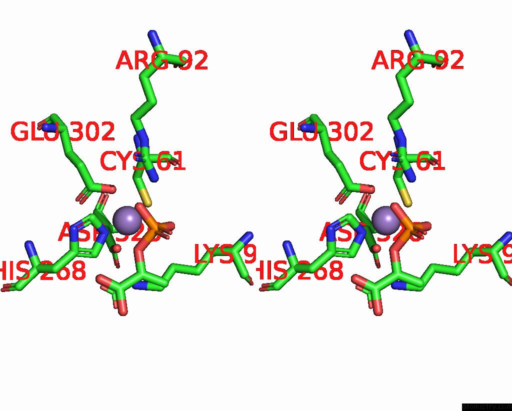
Manganese binding site 2 out of 8 in 1kfl
Go back to
Manganese binding site 2 out
of 8 in the Crystal Structure of Phenylalanine-Regulated 3-Deoxy-D- Arabino-Heptulosonate-7-Phosphate Synthase (Dahp Synthase) From E.Coli Complexed with MN2+, Pep, and Phe
Mono view
Stereo pair view
Mono view
Stereo pair view
A full contact list of Manganese with other atoms in the Mn binding
site number 2 of Crystal Structure of Phenylalanine-Regulated 3-Deoxy-D- Arabino-Heptulosonate-7-Phosphate Synthase (Dahp Synthase) From E.Coli Complexed with MN2+, Pep, and Phe within 5.0Å range:
|
Manganese binding site 3 out of 8 in 1kfl
Go back to
Manganese binding site 3 out
of 8 in the Crystal Structure of Phenylalanine-Regulated 3-Deoxy-D- Arabino-Heptulosonate-7-Phosphate Synthase (Dahp Synthase) From E.Coli Complexed with MN2+, Pep, and Phe
Mono view
Stereo pair view
Mono view
Stereo pair view
A full contact list of Manganese with other atoms in the Mn binding
site number 3 of Crystal Structure of Phenylalanine-Regulated 3-Deoxy-D- Arabino-Heptulosonate-7-Phosphate Synthase (Dahp Synthase) From E.Coli Complexed with MN2+, Pep, and Phe within 5.0Å range:
|
Manganese binding site 4 out of 8 in 1kfl
Go back to
Manganese binding site 4 out
of 8 in the Crystal Structure of Phenylalanine-Regulated 3-Deoxy-D- Arabino-Heptulosonate-7-Phosphate Synthase (Dahp Synthase) From E.Coli Complexed with MN2+, Pep, and Phe
Mono view
Stereo pair view
Mono view
Stereo pair view
A full contact list of Manganese with other atoms in the Mn binding
site number 4 of Crystal Structure of Phenylalanine-Regulated 3-Deoxy-D- Arabino-Heptulosonate-7-Phosphate Synthase (Dahp Synthase) From E.Coli Complexed with MN2+, Pep, and Phe within 5.0Å range:
|
Manganese binding site 5 out of 8 in 1kfl
Go back to
Manganese binding site 5 out
of 8 in the Crystal Structure of Phenylalanine-Regulated 3-Deoxy-D- Arabino-Heptulosonate-7-Phosphate Synthase (Dahp Synthase) From E.Coli Complexed with MN2+, Pep, and Phe
Mono view
Stereo pair view
Mono view
Stereo pair view
A full contact list of Manganese with other atoms in the Mn binding
site number 5 of Crystal Structure of Phenylalanine-Regulated 3-Deoxy-D- Arabino-Heptulosonate-7-Phosphate Synthase (Dahp Synthase) From E.Coli Complexed with MN2+, Pep, and Phe within 5.0Å range:
|
Manganese binding site 6 out of 8 in 1kfl
Go back to
Manganese binding site 6 out
of 8 in the Crystal Structure of Phenylalanine-Regulated 3-Deoxy-D- Arabino-Heptulosonate-7-Phosphate Synthase (Dahp Synthase) From E.Coli Complexed with MN2+, Pep, and Phe
Mono view
Stereo pair view
Mono view
Stereo pair view
A full contact list of Manganese with other atoms in the Mn binding
site number 6 of Crystal Structure of Phenylalanine-Regulated 3-Deoxy-D- Arabino-Heptulosonate-7-Phosphate Synthase (Dahp Synthase) From E.Coli Complexed with MN2+, Pep, and Phe within 5.0Å range:
|
Manganese binding site 7 out of 8 in 1kfl
Go back to
Manganese binding site 7 out
of 8 in the Crystal Structure of Phenylalanine-Regulated 3-Deoxy-D- Arabino-Heptulosonate-7-Phosphate Synthase (Dahp Synthase) From E.Coli Complexed with MN2+, Pep, and Phe
Mono view
Stereo pair view
Mono view
Stereo pair view
A full contact list of Manganese with other atoms in the Mn binding
site number 7 of Crystal Structure of Phenylalanine-Regulated 3-Deoxy-D- Arabino-Heptulosonate-7-Phosphate Synthase (Dahp Synthase) From E.Coli Complexed with MN2+, Pep, and Phe within 5.0Å range:
|
Manganese binding site 8 out of 8 in 1kfl
Go back to
Manganese binding site 8 out
of 8 in the Crystal Structure of Phenylalanine-Regulated 3-Deoxy-D- Arabino-Heptulosonate-7-Phosphate Synthase (Dahp Synthase) From E.Coli Complexed with MN2+, Pep, and Phe
Mono view
Stereo pair view
Mono view
Stereo pair view
A full contact list of Manganese with other atoms in the Mn binding
site number 8 of Crystal Structure of Phenylalanine-Regulated 3-Deoxy-D- Arabino-Heptulosonate-7-Phosphate Synthase (Dahp Synthase) From E.Coli Complexed with MN2+, Pep, and Phe within 5.0Å range:
|
Reference:
I.A.Shumilin,
C.Zhao,
R.Bauerle,
R.H.Kretsinger.
Allosteric Inhibition of 3-Deoxy-D-Arabino-Heptulosonate-7-Phosphate Synthase Alters the Coordination of Both Substrates. J.Mol.Biol. V. 320 1147 2002.
ISSN: ISSN 0022-2836
PubMed: 12126632
DOI: 10.1016/S0022-2836(02)00545-4
Page generated: Sat Oct 5 11:20:16 2024
ISSN: ISSN 0022-2836
PubMed: 12126632
DOI: 10.1016/S0022-2836(02)00545-4
Last articles
K in 4TM0K in 4TMZ
K in 4TLZ
K in 4TLX
K in 4RVO
K in 4RVN
K in 4TKX
K in 4RUM
K in 4RUF
K in 4RUE