Manganese »
PDB 1j54-1khb »
1jxn »
Manganese in PDB 1jxn: Crystal Structure of the Lectin I From Ulex Europaeus in Complex with the Methyl Glycoside of Alpha-L-Fucose
Protein crystallography data
The structure of Crystal Structure of the Lectin I From Ulex Europaeus in Complex with the Methyl Glycoside of Alpha-L-Fucose, PDB code: 1jxn
was solved by
G.F.Audette,
D.J.H.Olson,
A.R.S.Ross,
J.W.Quail,
L.T.J.Delbaere,
with X-Ray Crystallography technique. A brief refinement statistics is given in the table below:
| Resolution Low / High (Å) | 40.00 / 2.30 |
| Space group | P 1 21 1 |
| Cell size a, b, c (Å), α, β, γ (°) | 71.810, 69.000, 119.020, 90.00, 106.76, 90.00 |
| R / Rfree (%) | 20.2 / 28.9 |
Other elements in 1jxn:
The structure of Crystal Structure of the Lectin I From Ulex Europaeus in Complex with the Methyl Glycoside of Alpha-L-Fucose also contains other interesting chemical elements:
| Calcium | (Ca) | 4 atoms |
Manganese Binding Sites:
The binding sites of Manganese atom in the Crystal Structure of the Lectin I From Ulex Europaeus in Complex with the Methyl Glycoside of Alpha-L-Fucose
(pdb code 1jxn). This binding sites where shown within
5.0 Angstroms radius around Manganese atom.
In total 4 binding sites of Manganese where determined in the Crystal Structure of the Lectin I From Ulex Europaeus in Complex with the Methyl Glycoside of Alpha-L-Fucose, PDB code: 1jxn:
Jump to Manganese binding site number: 1; 2; 3; 4;
In total 4 binding sites of Manganese where determined in the Crystal Structure of the Lectin I From Ulex Europaeus in Complex with the Methyl Glycoside of Alpha-L-Fucose, PDB code: 1jxn:
Jump to Manganese binding site number: 1; 2; 3; 4;
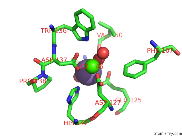
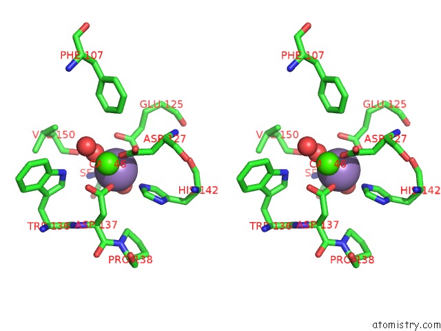
Manganese binding site 1 out of 4 in 1jxn
Go back to
Manganese binding site 1 out
of 4 in the Crystal Structure of the Lectin I From Ulex Europaeus in Complex with the Methyl Glycoside of Alpha-L-Fucose
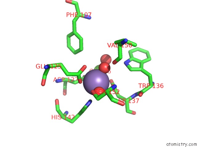
Mono view
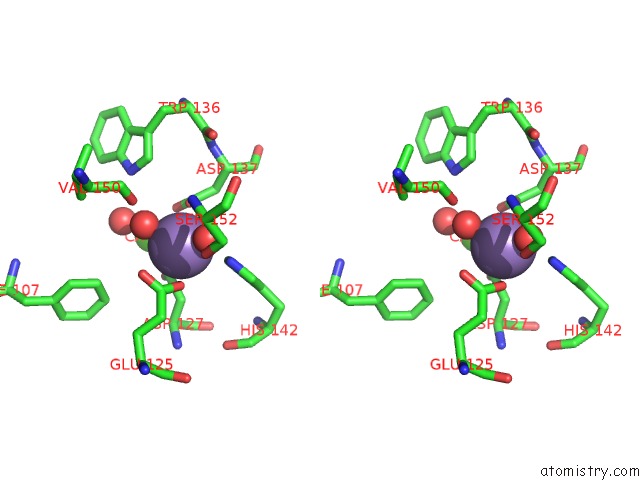
Stereo pair view
Mono view
Stereo pair view
A full contact list of Manganese with other atoms in the Mn binding
site number 1 of Crystal Structure of the Lectin I From Ulex Europaeus in Complex with the Methyl Glycoside of Alpha-L-Fucose within 5.0Å range:
|
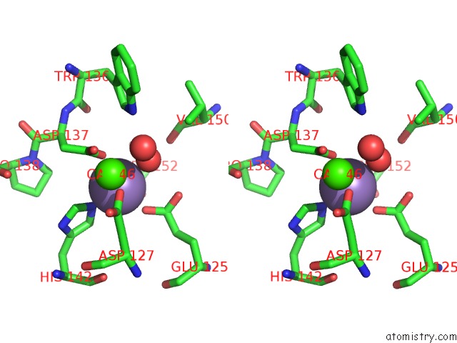
Manganese binding site 2 out of 4 in 1jxn
Go back to
Manganese binding site 2 out
of 4 in the Crystal Structure of the Lectin I From Ulex Europaeus in Complex with the Methyl Glycoside of Alpha-L-Fucose
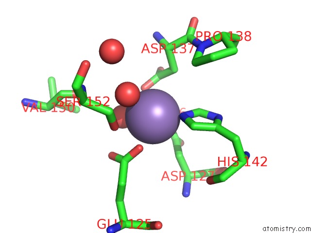
Mono view
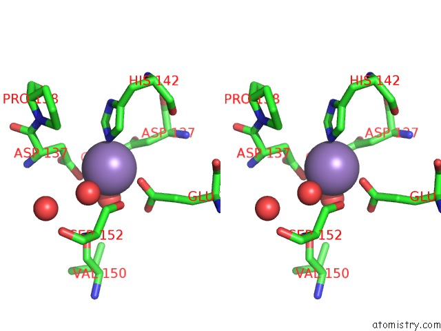
Stereo pair view
Mono view
Stereo pair view
A full contact list of Manganese with other atoms in the Mn binding
site number 2 of Crystal Structure of the Lectin I From Ulex Europaeus in Complex with the Methyl Glycoside of Alpha-L-Fucose within 5.0Å range:
|
Manganese binding site 3 out of 4 in 1jxn
Go back to
Manganese binding site 3 out
of 4 in the Crystal Structure of the Lectin I From Ulex Europaeus in Complex with the Methyl Glycoside of Alpha-L-Fucose
Mono view
Stereo pair view
Mono view
Stereo pair view
A full contact list of Manganese with other atoms in the Mn binding
site number 3 of Crystal Structure of the Lectin I From Ulex Europaeus in Complex with the Methyl Glycoside of Alpha-L-Fucose within 5.0Å range:
|
Manganese binding site 4 out of 4 in 1jxn
Go back to
Manganese binding site 4 out
of 4 in the Crystal Structure of the Lectin I From Ulex Europaeus in Complex with the Methyl Glycoside of Alpha-L-Fucose

Mono view
Stereo pair view
Mono view
Stereo pair view
A full contact list of Manganese with other atoms in the Mn binding
site number 4 of Crystal Structure of the Lectin I From Ulex Europaeus in Complex with the Methyl Glycoside of Alpha-L-Fucose within 5.0Å range:
|
Reference:
G.F.Audette,
D.J.H.Olson,
A.R.S.Ross,
J.W.Quail,
L.T.J.Delbaere.
Examination of the Structural Basis For O(H) Blood Group Specificity By Ulex Europaeus Lectin I Can.J.Chem. V. 80 1010 2002.
ISSN: ISSN 0008-4042
DOI: 10.1139/V02-134
Page generated: Sat Oct 5 11:15:48 2024
ISSN: ISSN 0008-4042
DOI: 10.1139/V02-134
Last articles
K in 4TMZK in 4TLZ
K in 4TLX
K in 4RVO
K in 4RVN
K in 4TKX
K in 4RUM
K in 4RUF
K in 4RUE
K in 4RGF