Manganese »
PDB 1j54-1khb »
1jk7 »
Manganese in PDB 1jk7: Crystal Structure of the Tumor-Promoter Okadaic Acid Bound to Protein Phosphatase-1
Enzymatic activity of Crystal Structure of the Tumor-Promoter Okadaic Acid Bound to Protein Phosphatase-1
All present enzymatic activity of Crystal Structure of the Tumor-Promoter Okadaic Acid Bound to Protein Phosphatase-1:
3.1.3.16;
3.1.3.16;
Protein crystallography data
The structure of Crystal Structure of the Tumor-Promoter Okadaic Acid Bound to Protein Phosphatase-1, PDB code: 1jk7
was solved by
J.T.Maynes,
K.S.Bateman,
M.M.Cherney,
A.K.Das,
M.N.James,
with X-Ray Crystallography technique. A brief refinement statistics is given in the table below:
| Resolution Low / High (Å) | 29.66 / 1.90 |
| Space group | P 42 21 2 |
| Cell size a, b, c (Å), α, β, γ (°) | 99.180, 99.180, 62.170, 90.00, 90.00, 90.00 |
| R / Rfree (%) | 19.9 / 22.5 |
Manganese Binding Sites:
The binding sites of Manganese atom in the Crystal Structure of the Tumor-Promoter Okadaic Acid Bound to Protein Phosphatase-1
(pdb code 1jk7). This binding sites where shown within
5.0 Angstroms radius around Manganese atom.
In total 2 binding sites of Manganese where determined in the Crystal Structure of the Tumor-Promoter Okadaic Acid Bound to Protein Phosphatase-1, PDB code: 1jk7:
Jump to Manganese binding site number: 1; 2;
In total 2 binding sites of Manganese where determined in the Crystal Structure of the Tumor-Promoter Okadaic Acid Bound to Protein Phosphatase-1, PDB code: 1jk7:
Jump to Manganese binding site number: 1; 2;
Manganese binding site 1 out of 2 in 1jk7
Go back to
Manganese binding site 1 out
of 2 in the Crystal Structure of the Tumor-Promoter Okadaic Acid Bound to Protein Phosphatase-1
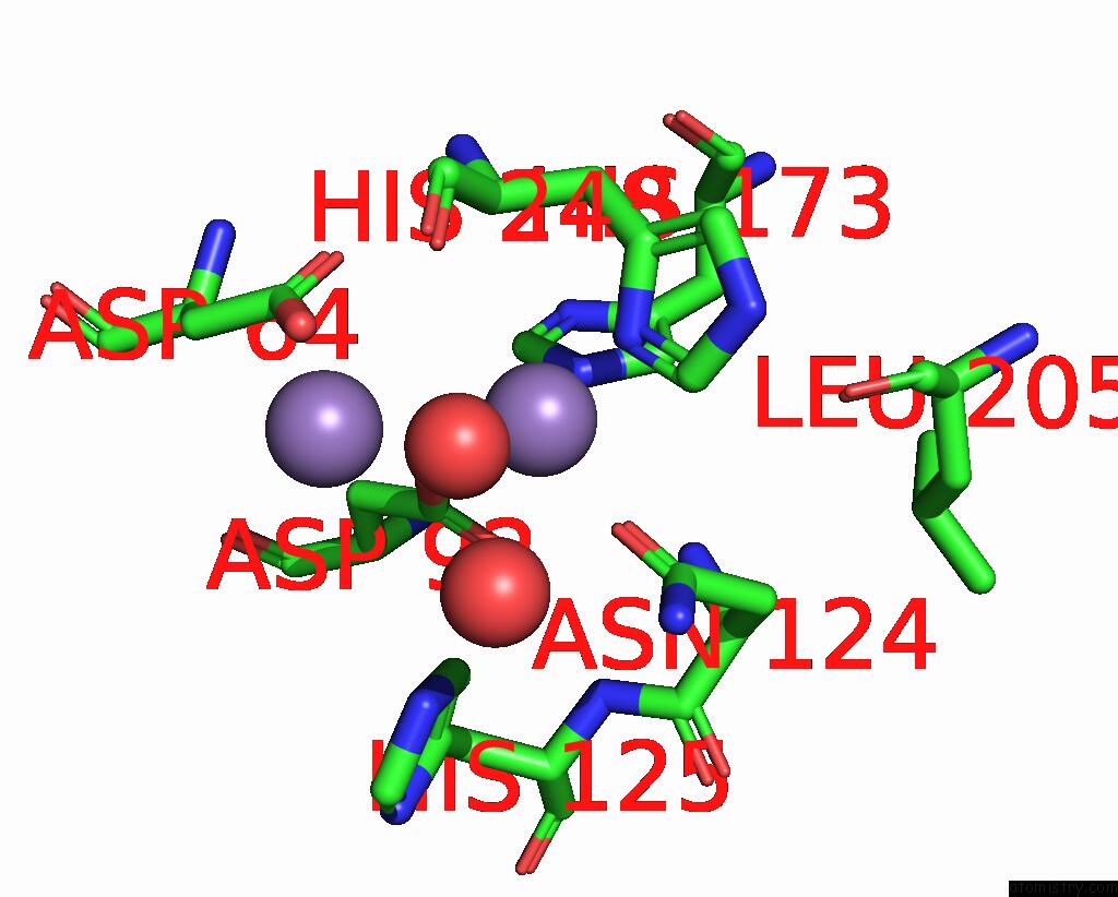
Mono view
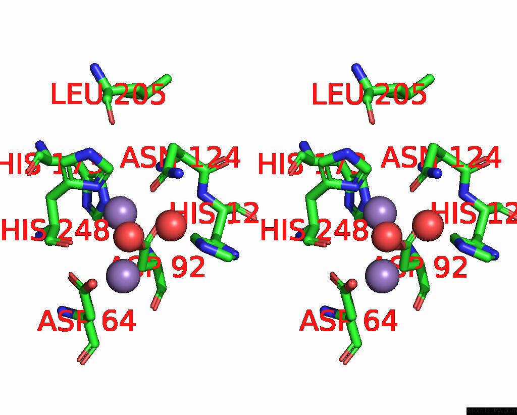
Stereo pair view
Mono view
Stereo pair view
A full contact list of Manganese with other atoms in the Mn binding
site number 1 of Crystal Structure of the Tumor-Promoter Okadaic Acid Bound to Protein Phosphatase-1 within 5.0Å range:
|
Manganese binding site 2 out of 2 in 1jk7
Go back to
Manganese binding site 2 out
of 2 in the Crystal Structure of the Tumor-Promoter Okadaic Acid Bound to Protein Phosphatase-1
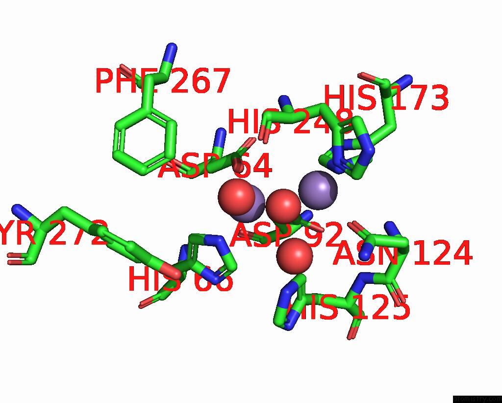
Mono view
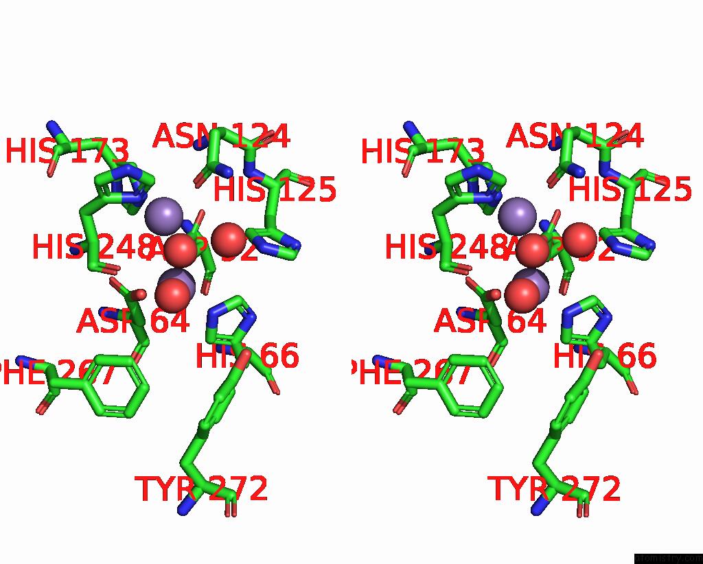
Stereo pair view
Mono view
Stereo pair view
A full contact list of Manganese with other atoms in the Mn binding
site number 2 of Crystal Structure of the Tumor-Promoter Okadaic Acid Bound to Protein Phosphatase-1 within 5.0Å range:
|
Reference:
J.T.Maynes,
K.S.Bateman,
M.M.Cherney,
A.K.Das,
H.A.Luu,
C.F.Holmes,
M.N.James.
Crystal Structure of the Tumor-Promoter Okadaic Acid Bound to Protein Phosphatase-1. J.Biol.Chem. V. 276 44078 2001.
ISSN: ISSN 0021-9258
PubMed: 11535607
DOI: 10.1074/JBC.M107656200
Page generated: Sat Oct 5 11:11:23 2024
ISSN: ISSN 0021-9258
PubMed: 11535607
DOI: 10.1074/JBC.M107656200
Last articles
Mg in 1JBZMg in 1JBW
Mg in 1JBV
Mg in 1JBK
Mg in 1JAX
Mg in 1JAH
Mg in 1J97
Mg in 1J9J
Mg in 1J8L
Mg in 1J7U