Manganese »
PDB 1g15-1hkd »
1gic »
Manganese in PDB 1gic: Concanavalin A Complexed with Methyl Alpha-D-Glucopyranoside
Protein crystallography data
The structure of Concanavalin A Complexed with Methyl Alpha-D-Glucopyranoside, PDB code: 1gic
was solved by
G.M.Bradbrook,
T.Gleichmann,
S.J.Harrop,
J.R.Helliwell,
J.Habash,
A.J.Kalb(Gilboa),
L.Tong,
T.C.M.Wan,
J.Yariv,
with X-Ray Crystallography technique. A brief refinement statistics is given in the table below:
| Resolution Low / High (Å) | 100.00 / 2.00 |
| Space group | I 21 3 |
| Cell size a, b, c (Å), α, β, γ (°) | 167.800, 167.800, 167.800, 90.00, 90.00, 90.00 |
| R / Rfree (%) | 17 / n/a |
Other elements in 1gic:
The structure of Concanavalin A Complexed with Methyl Alpha-D-Glucopyranoside also contains other interesting chemical elements:
| Calcium | (Ca) | 2 atoms |
Manganese Binding Sites:
The binding sites of Manganese atom in the Concanavalin A Complexed with Methyl Alpha-D-Glucopyranoside
(pdb code 1gic). This binding sites where shown within
5.0 Angstroms radius around Manganese atom.
In total 2 binding sites of Manganese where determined in the Concanavalin A Complexed with Methyl Alpha-D-Glucopyranoside, PDB code: 1gic:
Jump to Manganese binding site number: 1; 2;
In total 2 binding sites of Manganese where determined in the Concanavalin A Complexed with Methyl Alpha-D-Glucopyranoside, PDB code: 1gic:
Jump to Manganese binding site number: 1; 2;
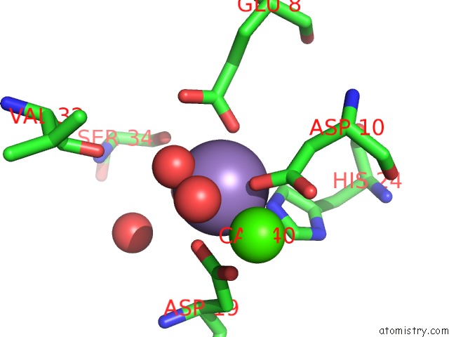
Manganese binding site 1 out of 2 in 1gic
Go back to
Manganese binding site 1 out
of 2 in the Concanavalin A Complexed with Methyl Alpha-D-Glucopyranoside
Mono view

Stereo pair view
Mono view
Stereo pair view
A full contact list of Manganese with other atoms in the Mn binding
site number 1 of Concanavalin A Complexed with Methyl Alpha-D-Glucopyranoside within 5.0Å range:
|
Manganese binding site 2 out of 2 in 1gic
Go back to
Manganese binding site 2 out
of 2 in the Concanavalin A Complexed with Methyl Alpha-D-Glucopyranoside

Mono view

Stereo pair view
Mono view
Stereo pair view
A full contact list of Manganese with other atoms in the Mn binding
site number 2 of Concanavalin A Complexed with Methyl Alpha-D-Glucopyranoside within 5.0Å range:
|
Reference:
S.J.Harrop,
J.R.Helliwell,
T.C.Wan,
A.J.Kalb,
L.Tong,
J.Yariv.
Structure Solution of A Cubic Crystal of Concanavalin A Complexed with Methyl Alpha-D-Glucopyranoside. Acta Crystallogr.,Sect.D V. 52 143 1996.
ISSN: ISSN 0907-4449
PubMed: 15299735
DOI: 10.1107/S0907444995008742
Page generated: Sat Oct 5 10:32:17 2024
ISSN: ISSN 0907-4449
PubMed: 15299735
DOI: 10.1107/S0907444995008742
Last articles
Fe in 6ZKPFe in 6ZKO
Fe in 6ZKM
Fe in 6ZKN
Fe in 6ZKL
Fe in 6ZKI
Fe in 6ZKK
Fe in 6ZKJ
Fe in 6ZKH
Fe in 6ZKG