Manganese »
PDB 8wq7-9c4d »
8z5b »
Manganese in PDB 8z5b: The X-Ray Crystal Structure of Multicopper Oxidase From Bacillus Freudenreichii
Enzymatic activity of The X-Ray Crystal Structure of Multicopper Oxidase From Bacillus Freudenreichii
All present enzymatic activity of The X-Ray Crystal Structure of Multicopper Oxidase From Bacillus Freudenreichii:
1.16.3.4;
1.16.3.4;
Protein crystallography data
The structure of The X-Ray Crystal Structure of Multicopper Oxidase From Bacillus Freudenreichii, PDB code: 8z5b
was solved by
H.Qian,
Y.Wang,
with X-Ray Crystallography technique. A brief refinement statistics is given in the table below:
| Resolution Low / High (Å) | 29.02 / 1.40 |
| Space group | P 41 21 2 |
| Cell size a, b, c (Å), α, β, γ (°) | 74.59, 74.59, 230.946, 90, 90, 90 |
| R / Rfree (%) | 15.8 / 17.3 |
Other elements in 8z5b:
The structure of The X-Ray Crystal Structure of Multicopper Oxidase From Bacillus Freudenreichii also contains other interesting chemical elements:
| Copper | (Cu) | 2 atoms |
Manganese Binding Sites:
The binding sites of Manganese atom in the The X-Ray Crystal Structure of Multicopper Oxidase From Bacillus Freudenreichii
(pdb code 8z5b). This binding sites where shown within
5.0 Angstroms radius around Manganese atom.
In total only one binding site of Manganese was determined in the The X-Ray Crystal Structure of Multicopper Oxidase From Bacillus Freudenreichii, PDB code: 8z5b:
In total only one binding site of Manganese was determined in the The X-Ray Crystal Structure of Multicopper Oxidase From Bacillus Freudenreichii, PDB code: 8z5b:
Manganese binding site 1 out of 1 in 8z5b
Go back to
Manganese binding site 1 out
of 1 in the The X-Ray Crystal Structure of Multicopper Oxidase From Bacillus Freudenreichii
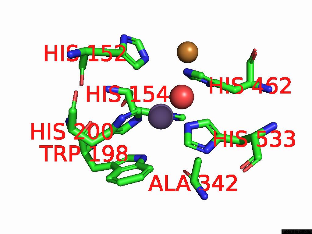
Mono view
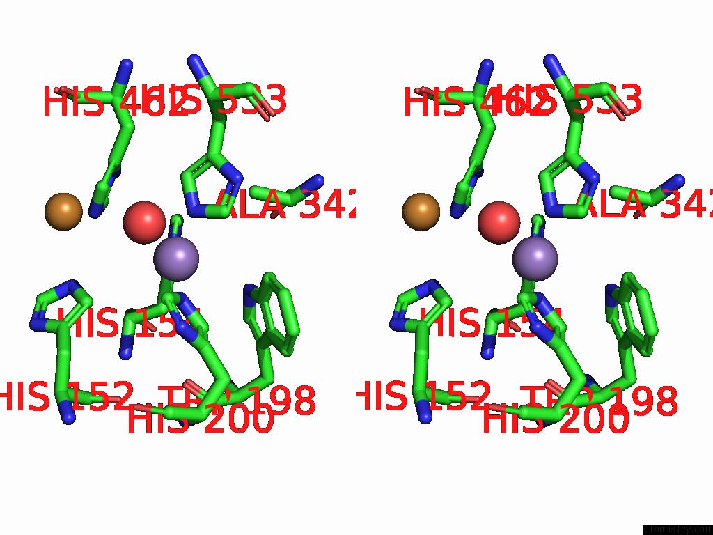
Stereo pair view
Mono view
Stereo pair view
A full contact list of Manganese with other atoms in the Mn binding
site number 1 of The X-Ray Crystal Structure of Multicopper Oxidase From Bacillus Freudenreichii within 5.0Å range:
|
Reference:
Y.Wang,
H.Qian.
The X-Ray Crystal Structure of Multicopper Oxidase From Bacillus Freudenreichii To Be Published.
Page generated: Tue Feb 25 11:29:39 2025
Last articles
Cl in 3EC8Cl in 3EBY
Cl in 3EBP
Cl in 3EAH
Cl in 3EAQ
Cl in 3E9Z
Cl in 3E8M
Cl in 3E9S
Cl in 3E82
Cl in 3E9L