Manganese »
PDB 8wq7-9c4d »
8yyk »
Manganese in PDB 8yyk: Structure of Rnase J2 Wild Type at Room Temperature
Protein crystallography data
The structure of Structure of Rnase J2 Wild Type at Room Temperature, PDB code: 8yyk
was solved by
A.K.Singh,
K.Chinnasamy,
B.Gopal,
with X-Ray Crystallography technique. A brief refinement statistics is given in the table below:
| Resolution Low / High (Å) | 58.60 / 3.20 |
| Space group | P 1 21 1 |
| Cell size a, b, c (Å), α, β, γ (°) | 65.304, 133.38, 117.751, 90, 92.02, 90 |
| R / Rfree (%) | 20.5 / 24.8 |
Manganese Binding Sites:
The binding sites of Manganese atom in the Structure of Rnase J2 Wild Type at Room Temperature
(pdb code 8yyk). This binding sites where shown within
5.0 Angstroms radius around Manganese atom.
In total 4 binding sites of Manganese where determined in the Structure of Rnase J2 Wild Type at Room Temperature, PDB code: 8yyk:
Jump to Manganese binding site number: 1; 2; 3; 4;
In total 4 binding sites of Manganese where determined in the Structure of Rnase J2 Wild Type at Room Temperature, PDB code: 8yyk:
Jump to Manganese binding site number: 1; 2; 3; 4;
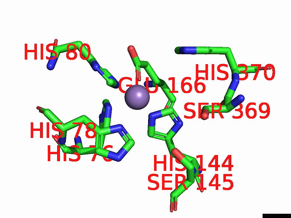
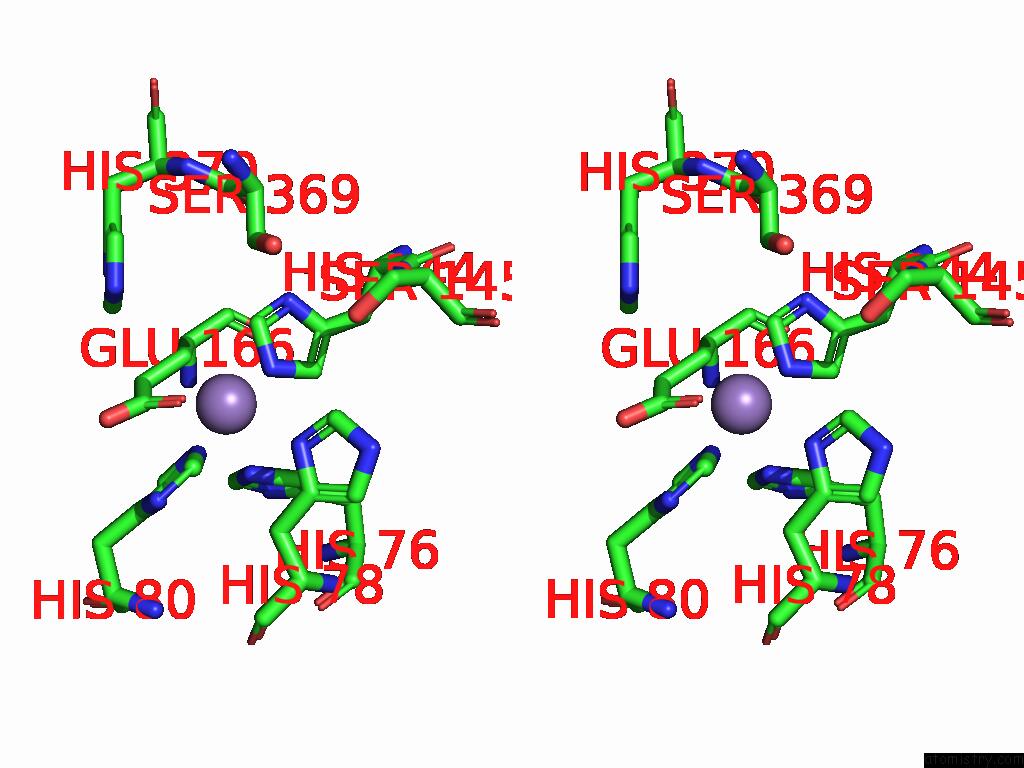
Manganese binding site 1 out of 4 in 8yyk
Go back to
Manganese binding site 1 out
of 4 in the Structure of Rnase J2 Wild Type at Room Temperature
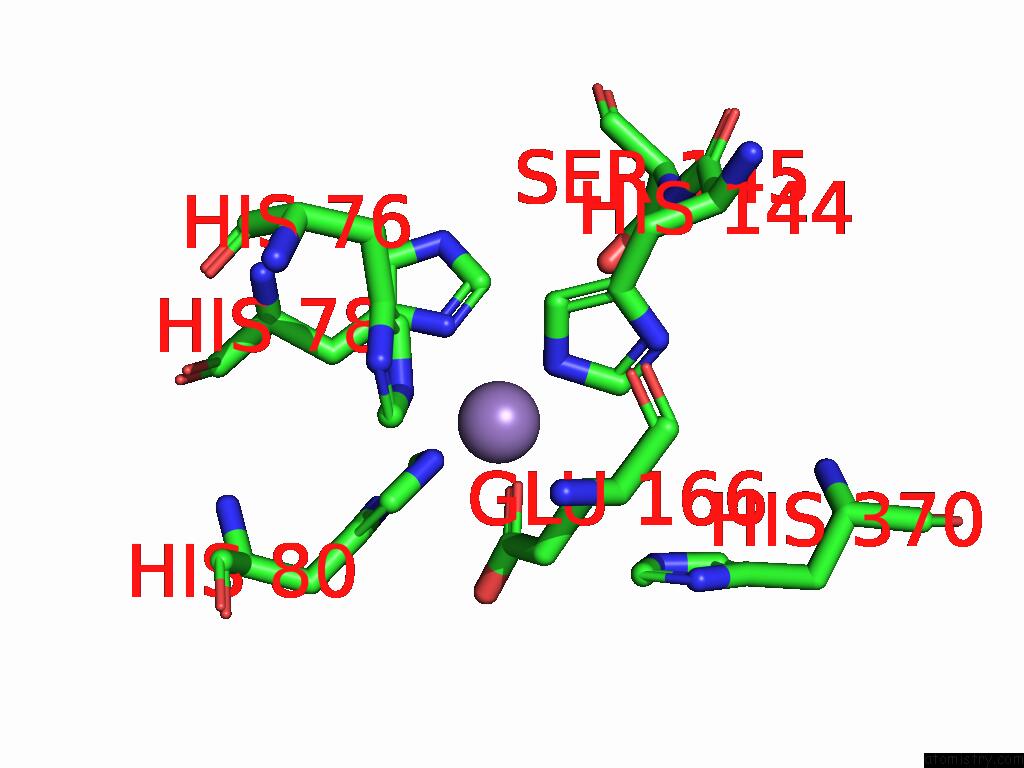
Mono view
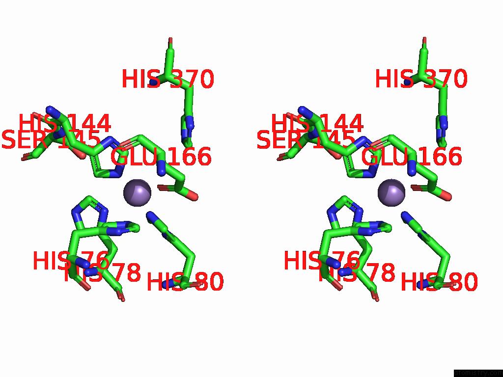
Stereo pair view
Mono view
Stereo pair view
A full contact list of Manganese with other atoms in the Mn binding
site number 1 of Structure of Rnase J2 Wild Type at Room Temperature within 5.0Å range:
|
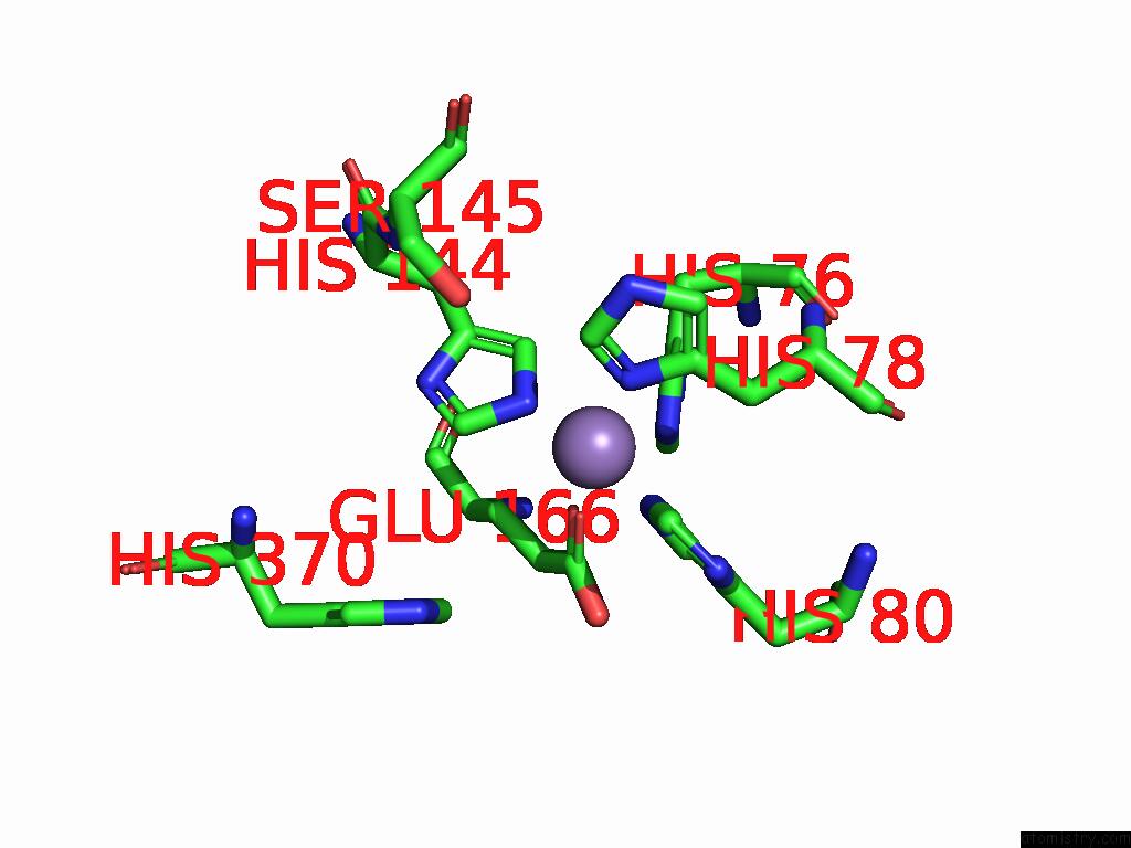
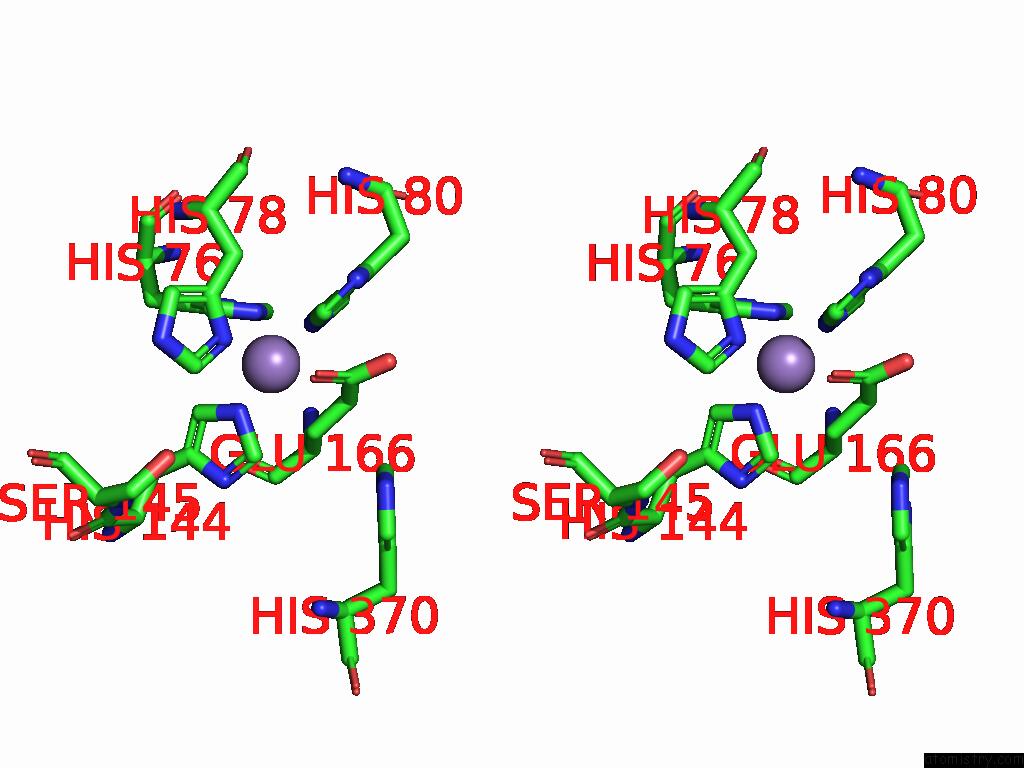
Manganese binding site 2 out of 4 in 8yyk
Go back to
Manganese binding site 2 out
of 4 in the Structure of Rnase J2 Wild Type at Room Temperature
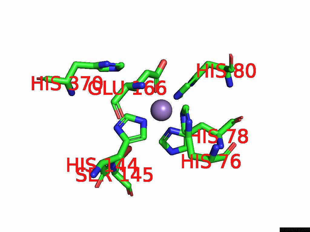
Mono view
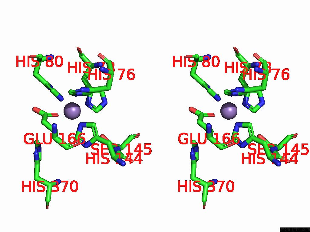
Stereo pair view
Mono view
Stereo pair view
A full contact list of Manganese with other atoms in the Mn binding
site number 2 of Structure of Rnase J2 Wild Type at Room Temperature within 5.0Å range:
|
Manganese binding site 3 out of 4 in 8yyk
Go back to
Manganese binding site 3 out
of 4 in the Structure of Rnase J2 Wild Type at Room Temperature
Mono view
Stereo pair view
Mono view
Stereo pair view
A full contact list of Manganese with other atoms in the Mn binding
site number 3 of Structure of Rnase J2 Wild Type at Room Temperature within 5.0Å range:
|
Manganese binding site 4 out of 4 in 8yyk
Go back to
Manganese binding site 4 out
of 4 in the Structure of Rnase J2 Wild Type at Room Temperature
Mono view
Stereo pair view
Mono view
Stereo pair view
A full contact list of Manganese with other atoms in the Mn binding
site number 4 of Structure of Rnase J2 Wild Type at Room Temperature within 5.0Å range:
|
Reference:
A.K.Singh,
K.Chinnasamy,
N.R.Pahelkar,
B.Gopal.
A Physicochemical Rationale For the Varied Catalytic Efficiency in Rnase J Paralogues. J.Biol.Chem. V. 301 08152 2024.
ISSN: ESSN 1083-351X
PubMed: 39742998
DOI: 10.1016/J.JBC.2024.108152
Page generated: Tue Feb 25 11:29:41 2025
ISSN: ESSN 1083-351X
PubMed: 39742998
DOI: 10.1016/J.JBC.2024.108152
Last articles
Fe in 2YXOFe in 2YRS
Fe in 2YXC
Fe in 2YNM
Fe in 2YVJ
Fe in 2YP1
Fe in 2YU2
Fe in 2YU1
Fe in 2YQB
Fe in 2YOO