Manganese »
PDB 8sln-8uwb »
8ujf »
Manganese in PDB 8ujf: X-Ray Crystal Structure of Toxoplasma Gondii Galnac-T3 in Complex with Udp-Galnac, MN2+, and MUC5AC-3,13
Enzymatic activity of X-Ray Crystal Structure of Toxoplasma Gondii Galnac-T3 in Complex with Udp-Galnac, MN2+, and MUC5AC-3,13
All present enzymatic activity of X-Ray Crystal Structure of Toxoplasma Gondii Galnac-T3 in Complex with Udp-Galnac, MN2+, and MUC5AC-3,13:
2.4.1.41;
2.4.1.41;
Protein crystallography data
The structure of X-Ray Crystal Structure of Toxoplasma Gondii Galnac-T3 in Complex with Udp-Galnac, MN2+, and MUC5AC-3,13, PDB code: 8ujf
was solved by
P.Kumar,
N.L.Samara,
with X-Ray Crystallography technique. A brief refinement statistics is given in the table below:
| Resolution Low / High (Å) | 20.01 / 2.87 |
| Space group | P 21 21 21 |
| Cell size a, b, c (Å), α, β, γ (°) | 66.233, 123.693, 165.724, 90, 90, 90 |
| R / Rfree (%) | 21.7 / 27.4 |
Manganese Binding Sites:
The binding sites of Manganese atom in the X-Ray Crystal Structure of Toxoplasma Gondii Galnac-T3 in Complex with Udp-Galnac, MN2+, and MUC5AC-3,13
(pdb code 8ujf). This binding sites where shown within
5.0 Angstroms radius around Manganese atom.
In total 4 binding sites of Manganese where determined in the X-Ray Crystal Structure of Toxoplasma Gondii Galnac-T3 in Complex with Udp-Galnac, MN2+, and MUC5AC-3,13, PDB code: 8ujf:
Jump to Manganese binding site number: 1; 2; 3; 4;
In total 4 binding sites of Manganese where determined in the X-Ray Crystal Structure of Toxoplasma Gondii Galnac-T3 in Complex with Udp-Galnac, MN2+, and MUC5AC-3,13, PDB code: 8ujf:
Jump to Manganese binding site number: 1; 2; 3; 4;
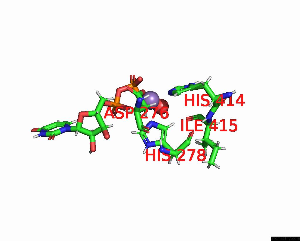
Manganese binding site 1 out of 4 in 8ujf
Go back to
Manganese binding site 1 out
of 4 in the X-Ray Crystal Structure of Toxoplasma Gondii Galnac-T3 in Complex with Udp-Galnac, MN2+, and MUC5AC-3,13
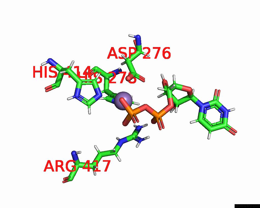
Mono view
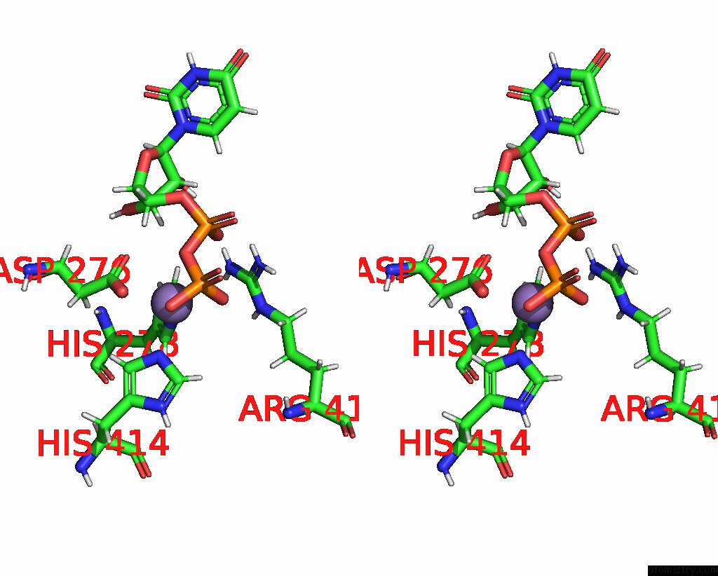
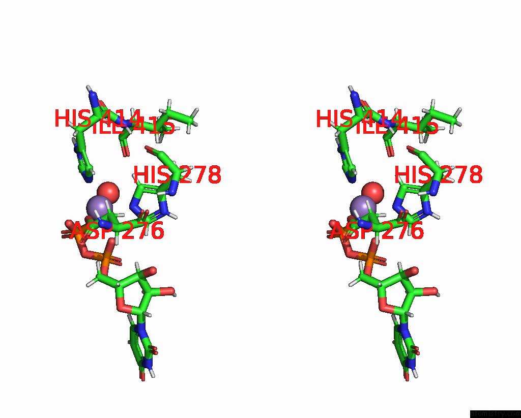
Stereo pair view
Mono view
Stereo pair view
A full contact list of Manganese with other atoms in the Mn binding
site number 1 of X-Ray Crystal Structure of Toxoplasma Gondii Galnac-T3 in Complex with Udp-Galnac, MN2+, and MUC5AC-3,13 within 5.0Å range:
|
Manganese binding site 2 out of 4 in 8ujf
Go back to
Manganese binding site 2 out
of 4 in the X-Ray Crystal Structure of Toxoplasma Gondii Galnac-T3 in Complex with Udp-Galnac, MN2+, and MUC5AC-3,13
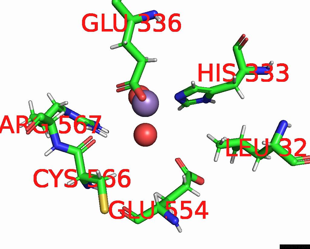
Mono view
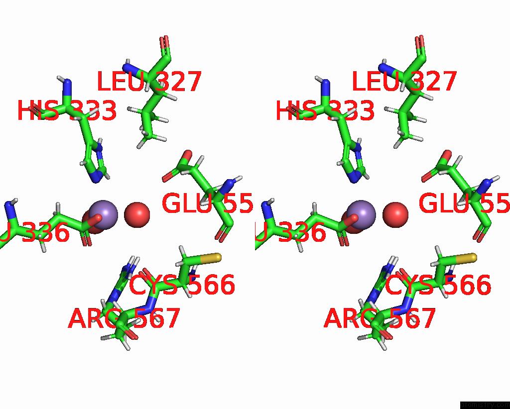
Stereo pair view
Mono view
Stereo pair view
A full contact list of Manganese with other atoms in the Mn binding
site number 2 of X-Ray Crystal Structure of Toxoplasma Gondii Galnac-T3 in Complex with Udp-Galnac, MN2+, and MUC5AC-3,13 within 5.0Å range:
|
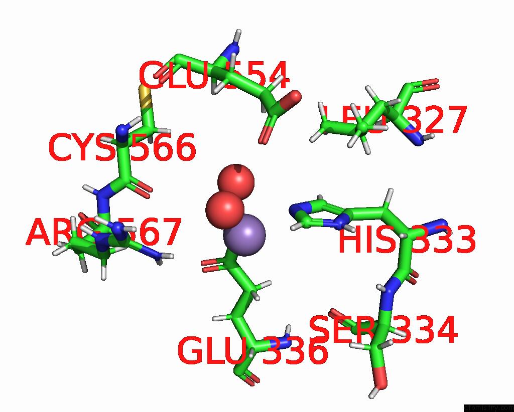
Manganese binding site 3 out of 4 in 8ujf
Go back to
Manganese binding site 3 out
of 4 in the X-Ray Crystal Structure of Toxoplasma Gondii Galnac-T3 in Complex with Udp-Galnac, MN2+, and MUC5AC-3,13
Mono view
Stereo pair view
Mono view
Stereo pair view
A full contact list of Manganese with other atoms in the Mn binding
site number 3 of X-Ray Crystal Structure of Toxoplasma Gondii Galnac-T3 in Complex with Udp-Galnac, MN2+, and MUC5AC-3,13 within 5.0Å range:
|
Manganese binding site 4 out of 4 in 8ujf
Go back to
Manganese binding site 4 out
of 4 in the X-Ray Crystal Structure of Toxoplasma Gondii Galnac-T3 in Complex with Udp-Galnac, MN2+, and MUC5AC-3,13
Mono view
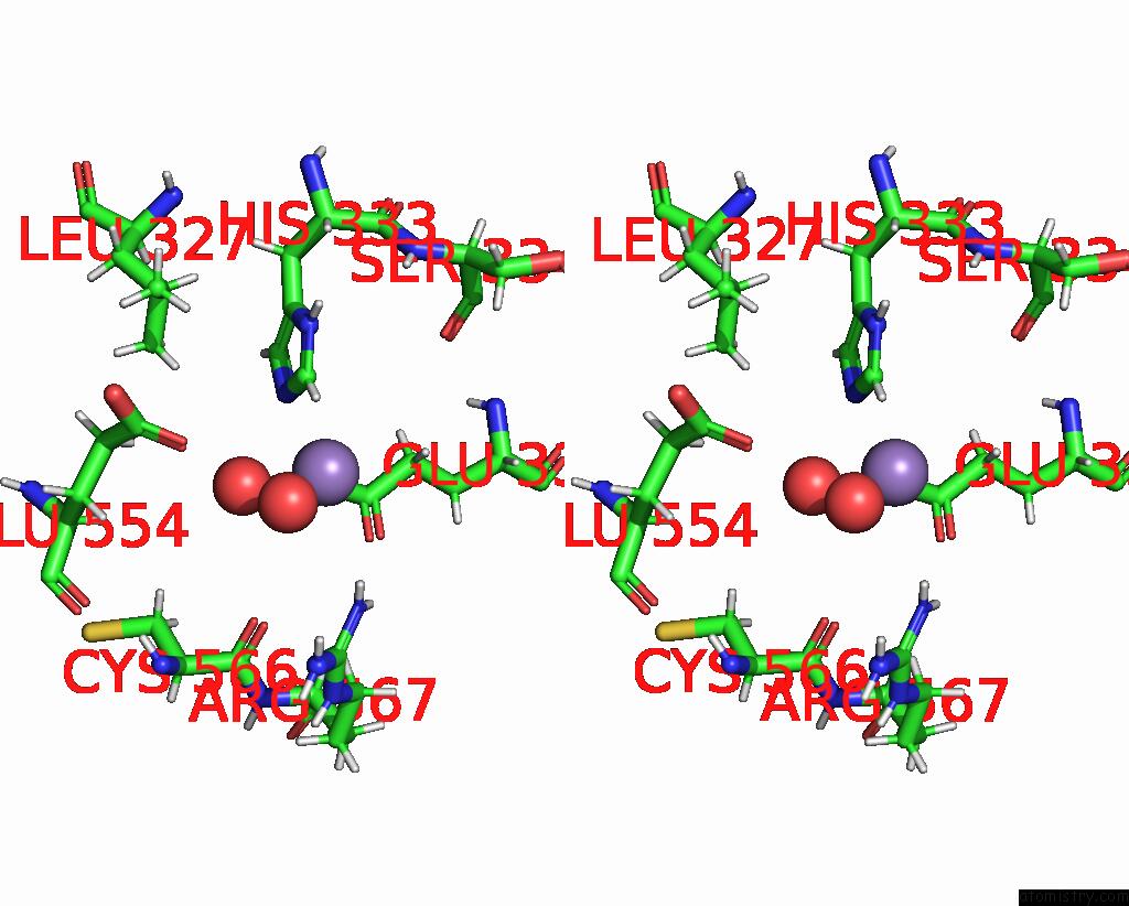
Stereo pair view
Mono view
Stereo pair view
A full contact list of Manganese with other atoms in the Mn binding
site number 4 of X-Ray Crystal Structure of Toxoplasma Gondii Galnac-T3 in Complex with Udp-Galnac, MN2+, and MUC5AC-3,13 within 5.0Å range:
|
Reference:
P.Kumar,
T.Tomita,
T.A.Gerken,
C.J.Ballard,
Y.S.Lee,
L.M.Weiss,
N.L.Samara.
A Toxoplasma Gondii O-Glycosyltransferase That Modulates Bradyzoite Cyst Wall Rigidity Is Distinct From Host Homologues Nat Commun V. 15 3792 2024.
ISSN: ESSN 2041-1723
DOI: 10.1038/S41467-024-48253-W
Page generated: Sun Oct 6 14:00:52 2024
ISSN: ESSN 2041-1723
DOI: 10.1038/S41467-024-48253-W
Last articles
Zn in 9MJ5Zn in 9HNW
Zn in 9G0L
Zn in 9FNE
Zn in 9DZN
Zn in 9E0I
Zn in 9D32
Zn in 9DAK
Zn in 8ZXC
Zn in 8ZUF