Manganese »
PDB 8sln-8uwb »
8tjc »
Manganese in PDB 8tjc: Structure of Human Beta 1,3-N-Acetylglucosaminyltransferase 2 with Compound 8A
Enzymatic activity of Structure of Human Beta 1,3-N-Acetylglucosaminyltransferase 2 with Compound 8A
All present enzymatic activity of Structure of Human Beta 1,3-N-Acetylglucosaminyltransferase 2 with Compound 8A:
2.4.1.149;
2.4.1.149;
Protein crystallography data
The structure of Structure of Human Beta 1,3-N-Acetylglucosaminyltransferase 2 with Compound 8A, PDB code: 8tjc
was solved by
A.Sudom,
X.Min,
with X-Ray Crystallography technique. A brief refinement statistics is given in the table below:
| Resolution Low / High (Å) | 47.01 / 2.20 |
| Space group | P 21 21 21 |
| Cell size a, b, c (Å), α, β, γ (°) | 89.41, 93.876, 205.066, 90, 90, 90 |
| R / Rfree (%) | 22.7 / 27.5 |
Manganese Binding Sites:
The binding sites of Manganese atom in the Structure of Human Beta 1,3-N-Acetylglucosaminyltransferase 2 with Compound 8A
(pdb code 8tjc). This binding sites where shown within
5.0 Angstroms radius around Manganese atom.
In total 4 binding sites of Manganese where determined in the Structure of Human Beta 1,3-N-Acetylglucosaminyltransferase 2 with Compound 8A, PDB code: 8tjc:
Jump to Manganese binding site number: 1; 2; 3; 4;
In total 4 binding sites of Manganese where determined in the Structure of Human Beta 1,3-N-Acetylglucosaminyltransferase 2 with Compound 8A, PDB code: 8tjc:
Jump to Manganese binding site number: 1; 2; 3; 4;
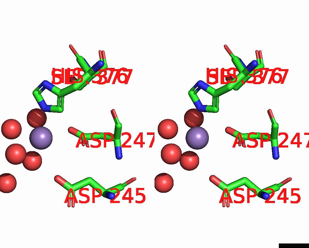
Manganese binding site 1 out of 4 in 8tjc
Go back to
Manganese binding site 1 out
of 4 in the Structure of Human Beta 1,3-N-Acetylglucosaminyltransferase 2 with Compound 8A

Mono view
Stereo pair view
Mono view
Stereo pair view
A full contact list of Manganese with other atoms in the Mn binding
site number 1 of Structure of Human Beta 1,3-N-Acetylglucosaminyltransferase 2 with Compound 8A within 5.0Å range:
|
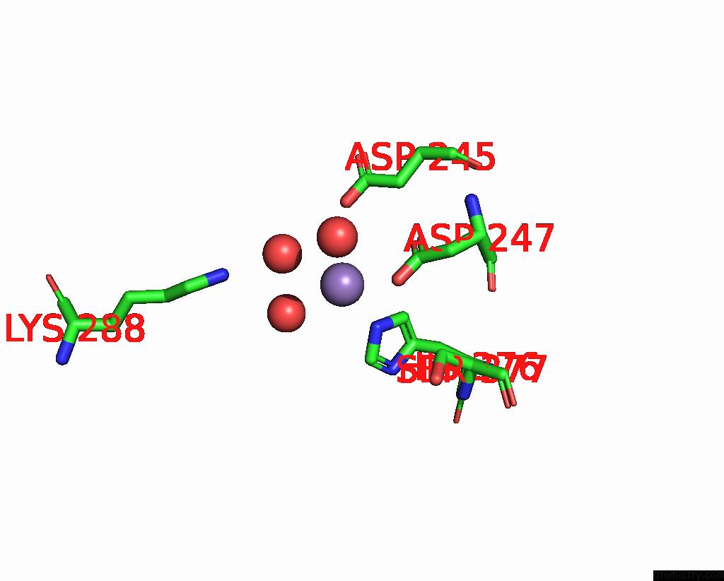
Manganese binding site 2 out of 4 in 8tjc
Go back to
Manganese binding site 2 out
of 4 in the Structure of Human Beta 1,3-N-Acetylglucosaminyltransferase 2 with Compound 8A

Mono view

Stereo pair view
Mono view
Stereo pair view
A full contact list of Manganese with other atoms in the Mn binding
site number 2 of Structure of Human Beta 1,3-N-Acetylglucosaminyltransferase 2 with Compound 8A within 5.0Å range:
|
Manganese binding site 3 out of 4 in 8tjc
Go back to
Manganese binding site 3 out
of 4 in the Structure of Human Beta 1,3-N-Acetylglucosaminyltransferase 2 with Compound 8A
Mono view

Stereo pair view
Mono view
Stereo pair view
A full contact list of Manganese with other atoms in the Mn binding
site number 3 of Structure of Human Beta 1,3-N-Acetylglucosaminyltransferase 2 with Compound 8A within 5.0Å range:
|
Manganese binding site 4 out of 4 in 8tjc
Go back to
Manganese binding site 4 out
of 4 in the Structure of Human Beta 1,3-N-Acetylglucosaminyltransferase 2 with Compound 8A

Mono view

Stereo pair view
Mono view
Stereo pair view
A full contact list of Manganese with other atoms in the Mn binding
site number 4 of Structure of Human Beta 1,3-N-Acetylglucosaminyltransferase 2 with Compound 8A within 5.0Å range:
|
Reference:
J.J.Jackson,
A.C.Siegmund,
W.J.Bai,
A.B.Reed,
A.B.Birkholz,
I.D.G.Campuzano,
A.Crequer-Grandhomme,
R.Hu,
R.V.Modak,
A.Sudom,
N.Javier,
C.Sanders,
M.C.Lo,
F.Xie,
V.J.Cee,
P.Manzanillo,
J.G.Allen.
Imidazolone As An Amide Bioisostere in the Development of Beta-1,3- N -Acetylglucosaminyltransferase 2 (B3GNT2) Inhibitors. J.Med.Chem. 2023.
ISSN: ISSN 0022-2623
PubMed: 37988652
DOI: 10.1021/ACS.JMEDCHEM.3C01517
Page generated: Sun Oct 6 13:57:16 2024
ISSN: ISSN 0022-2623
PubMed: 37988652
DOI: 10.1021/ACS.JMEDCHEM.3C01517
Last articles
Ca in 5OEOCa in 5OF0
Ca in 5OC9
Ca in 5ODC
Ca in 5O8N
Ca in 5O8X
Ca in 5O32
Ca in 5O76
Ca in 5O8L
Ca in 5O7W