Manganese »
PDB 8sln-8uwb »
8tf0 »
Manganese in PDB 8tf0: Crystal Structure of GRP94 N-Terminal Domain Bound to the Purine Inhibitor Pu-H36
Protein crystallography data
The structure of Crystal Structure of GRP94 N-Terminal Domain Bound to the Purine Inhibitor Pu-H36, PDB code: 8tf0
was solved by
N.L.S.Que,
D.T.Gewirth,
with X-Ray Crystallography technique. A brief refinement statistics is given in the table below:
| Resolution Low / High (Å) | 36.89 / 2.79 |
| Space group | P 21 21 21 |
| Cell size a, b, c (Å), α, β, γ (°) | 96.282, 104.392, 172.284, 90, 90, 90 |
| R / Rfree (%) | 22.8 / 26 |
Other elements in 8tf0:
The structure of Crystal Structure of GRP94 N-Terminal Domain Bound to the Purine Inhibitor Pu-H36 also contains other interesting chemical elements:
| Magnesium | (Mg) | 11 atoms |
Manganese Binding Sites:
The binding sites of Manganese atom in the Crystal Structure of GRP94 N-Terminal Domain Bound to the Purine Inhibitor Pu-H36
(pdb code 8tf0). This binding sites where shown within
5.0 Angstroms radius around Manganese atom.
In total only one binding site of Manganese was determined in the Crystal Structure of GRP94 N-Terminal Domain Bound to the Purine Inhibitor Pu-H36, PDB code: 8tf0:
In total only one binding site of Manganese was determined in the Crystal Structure of GRP94 N-Terminal Domain Bound to the Purine Inhibitor Pu-H36, PDB code: 8tf0:
Manganese binding site 1 out of 1 in 8tf0
Go back to
Manganese binding site 1 out
of 1 in the Crystal Structure of GRP94 N-Terminal Domain Bound to the Purine Inhibitor Pu-H36
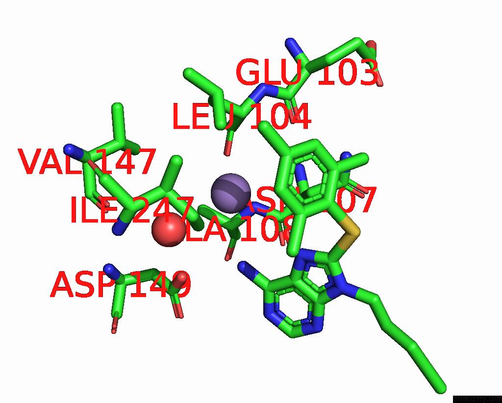
Mono view
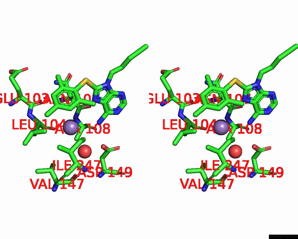
Stereo pair view
Mono view
Stereo pair view
A full contact list of Manganese with other atoms in the Mn binding
site number 1 of Crystal Structure of GRP94 N-Terminal Domain Bound to the Purine Inhibitor Pu-H36 within 5.0Å range:
|
Reference:
N.L.S.Que,
P.M.Seidler,
W.J.Aw,
G.Chiosis,
D.T.Gewirth.
Selective Inhibition of HSP90 Paralogs: Uncovering the Role of Helix 1 in GRP94-Selective Ligand Binding. Proteins 2024.
ISSN: ESSN 1097-0134
PubMed: 39473058
DOI: 10.1002/PROT.26756
Page generated: Wed Nov 13 12:56:10 2024
ISSN: ESSN 1097-0134
PubMed: 39473058
DOI: 10.1002/PROT.26756
Last articles
Zn in 9MJ5Zn in 9HNW
Zn in 9G0L
Zn in 9FNE
Zn in 9DZN
Zn in 9E0I
Zn in 9D32
Zn in 9DAK
Zn in 8ZXC
Zn in 8ZUF