Manganese »
PDB 8q3z-8slm »
8qzj »
Manganese in PDB 8qzj: Crystal Structure of Pptt-Adp From Mycobacterium Tuberculosis
Protein crystallography data
The structure of Crystal Structure of Pptt-Adp From Mycobacterium Tuberculosis, PDB code: 8qzj
was solved by
S.Gavalda,
L.Mourey,
J.D.Pedelacq,
with X-Ray Crystallography technique. A brief refinement statistics is given in the table below:
| Resolution Low / High (Å) | 47.97 / 2.00 |
| Space group | P 2 21 21 |
| Cell size a, b, c (Å), α, β, γ (°) | 65.86, 68.792, 210.063, 90, 90, 90 |
| R / Rfree (%) | 21.9 / 26.8 |
Manganese Binding Sites:
The binding sites of Manganese atom in the Crystal Structure of Pptt-Adp From Mycobacterium Tuberculosis
(pdb code 8qzj). This binding sites where shown within
5.0 Angstroms radius around Manganese atom.
In total 8 binding sites of Manganese where determined in the Crystal Structure of Pptt-Adp From Mycobacterium Tuberculosis, PDB code: 8qzj:
Jump to Manganese binding site number: 1; 2; 3; 4; 5; 6; 7; 8;
In total 8 binding sites of Manganese where determined in the Crystal Structure of Pptt-Adp From Mycobacterium Tuberculosis, PDB code: 8qzj:
Jump to Manganese binding site number: 1; 2; 3; 4; 5; 6; 7; 8;
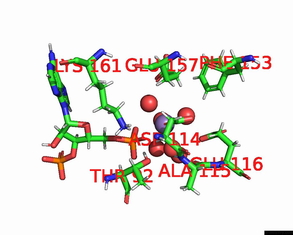
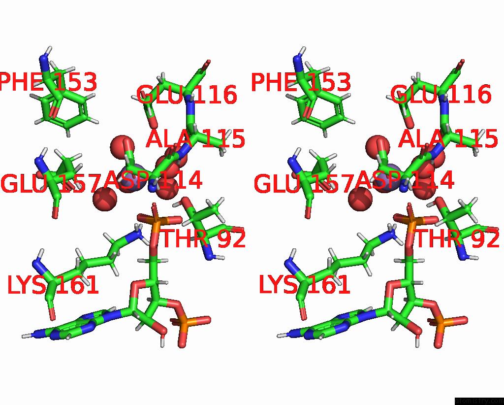
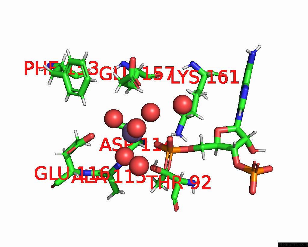
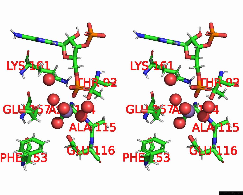
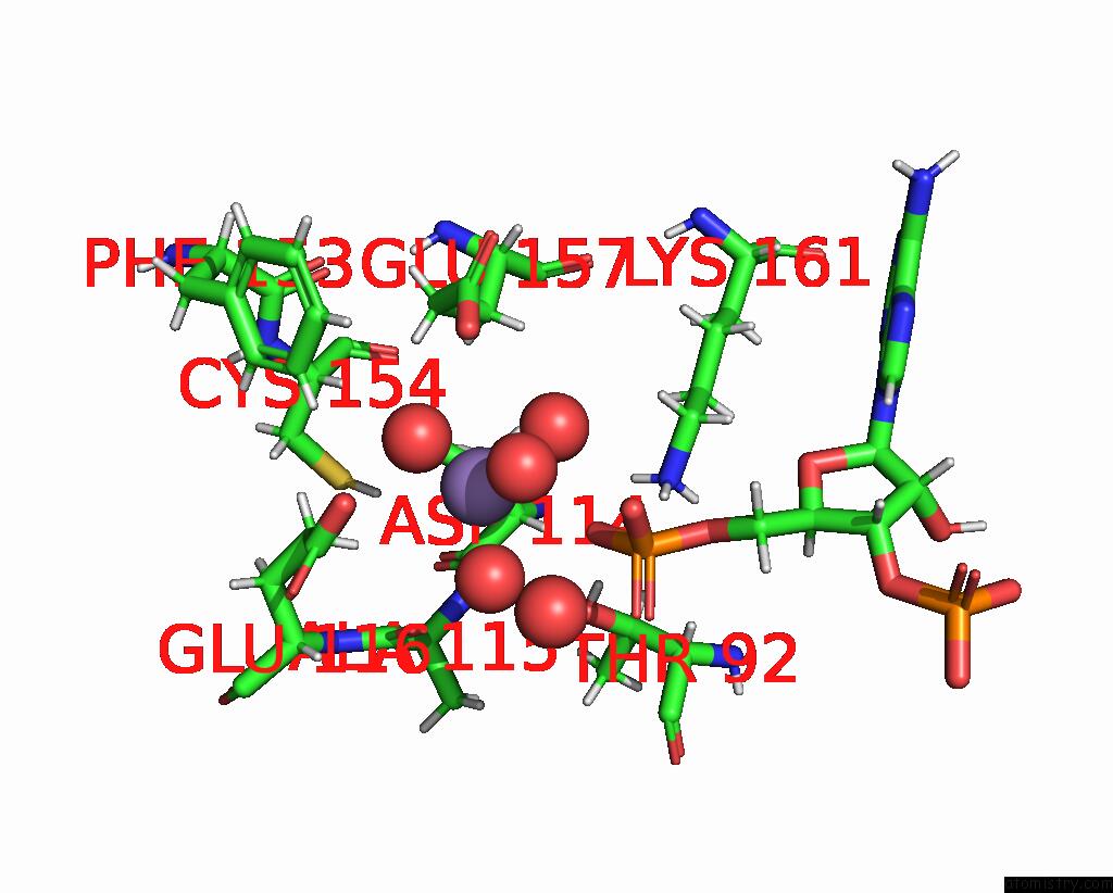
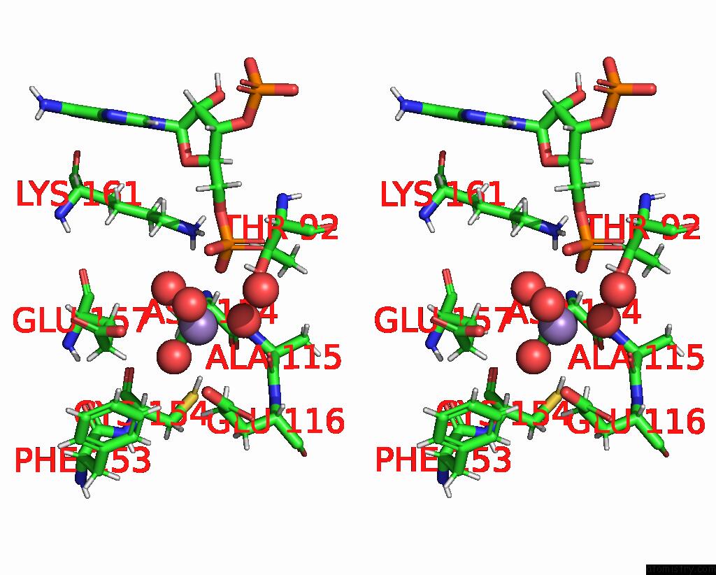
Manganese binding site 1 out of 8 in 8qzj
Go back to
Manganese binding site 1 out
of 8 in the Crystal Structure of Pptt-Adp From Mycobacterium Tuberculosis
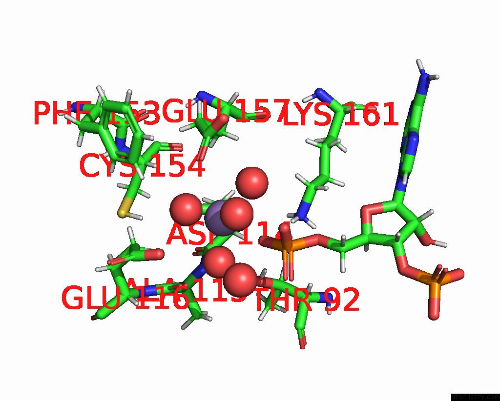
Mono view
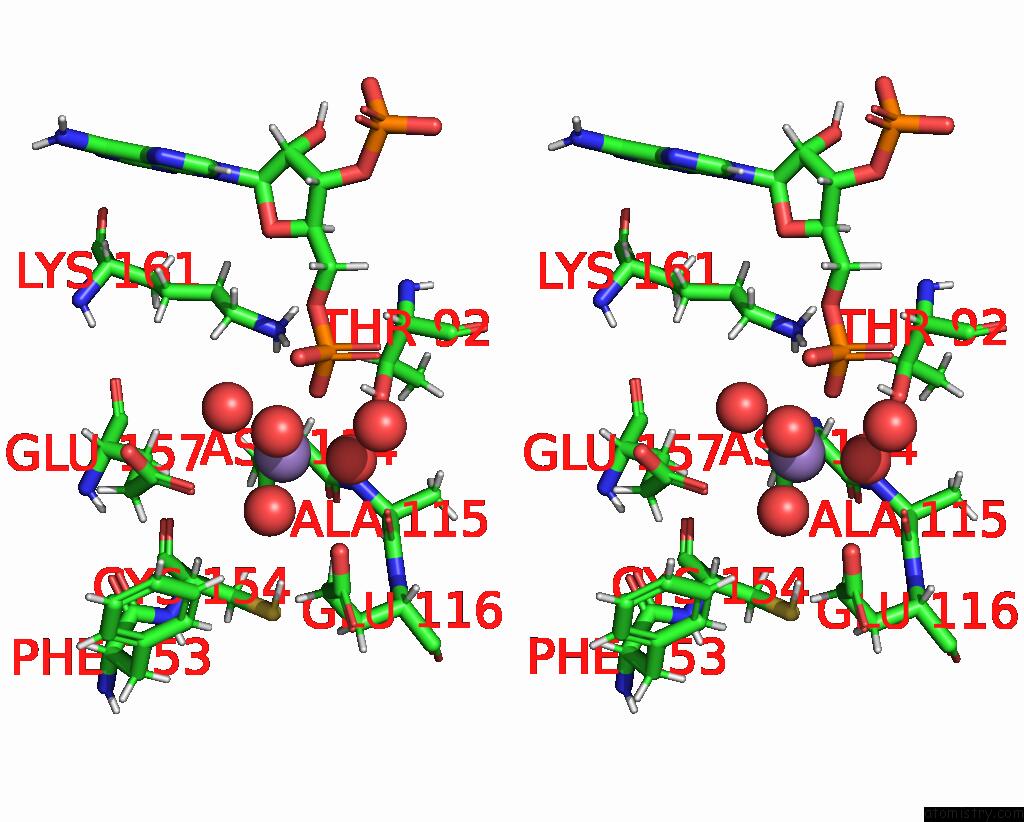
Stereo pair view
Mono view
Stereo pair view
A full contact list of Manganese with other atoms in the Mn binding
site number 1 of Crystal Structure of Pptt-Adp From Mycobacterium Tuberculosis within 5.0Å range:
|
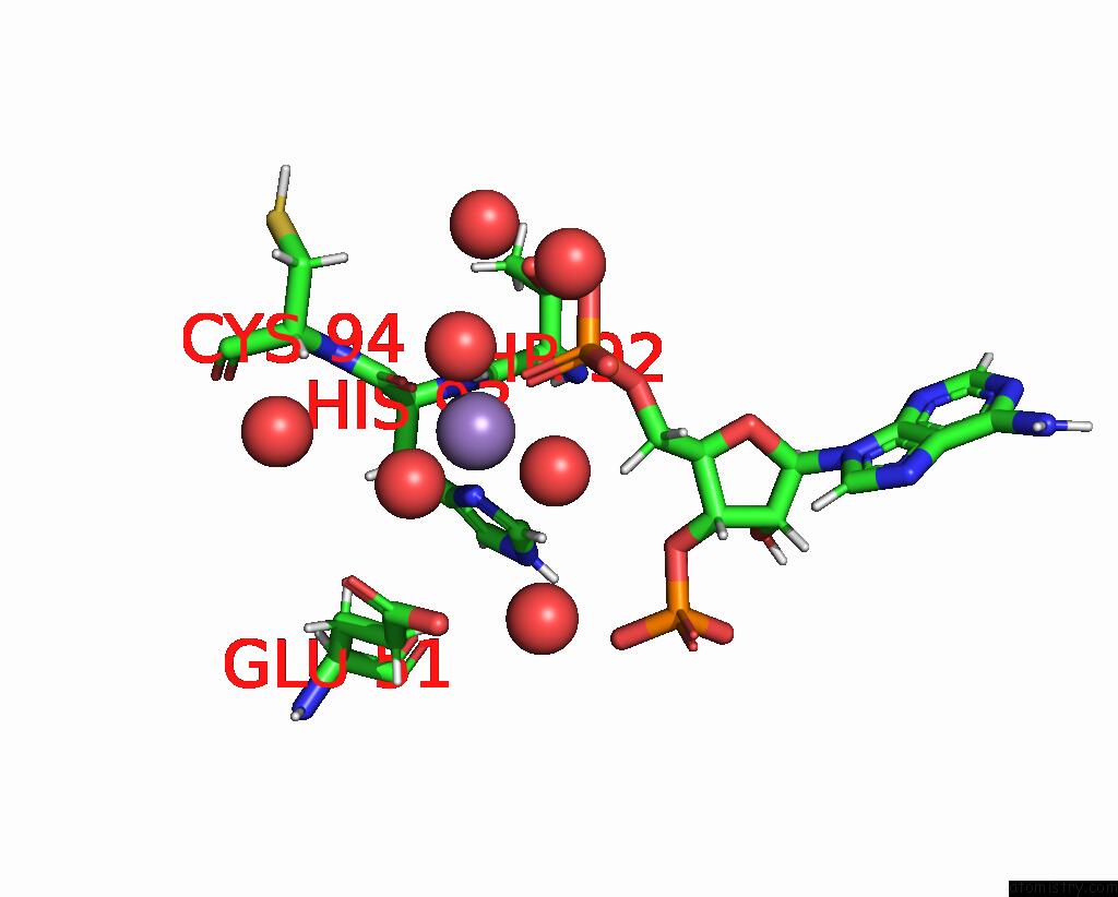
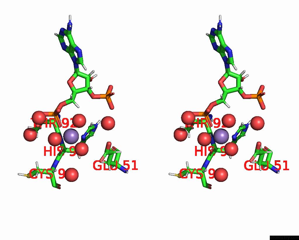
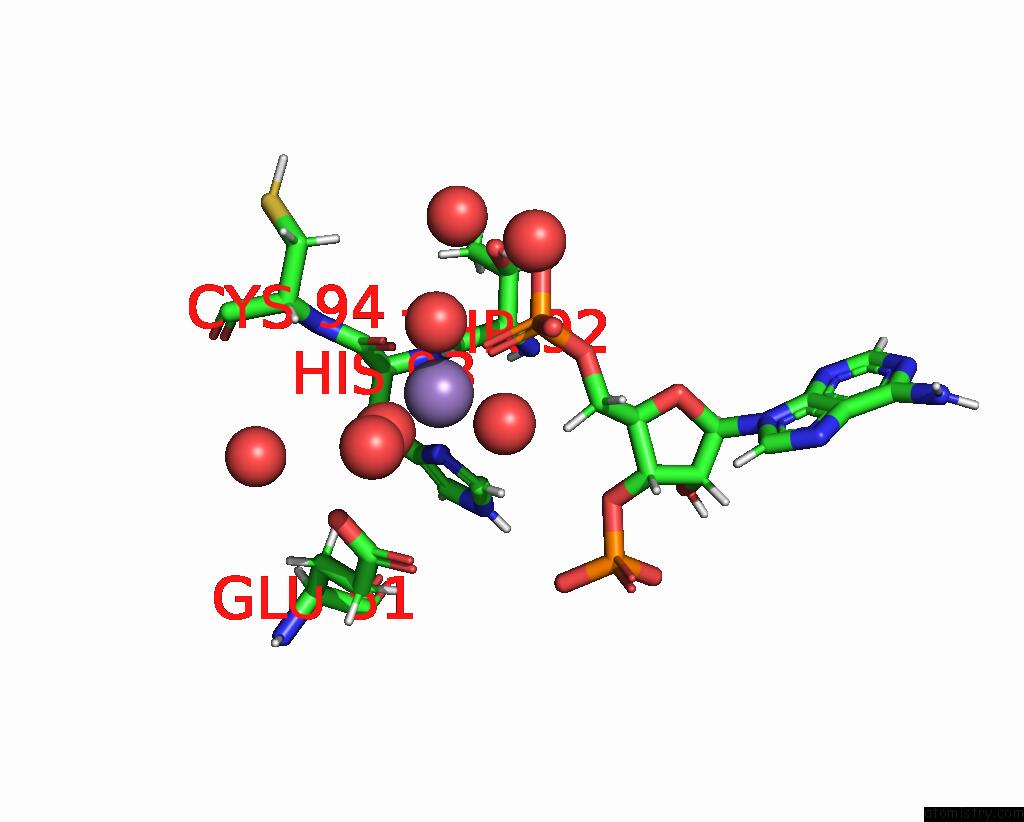
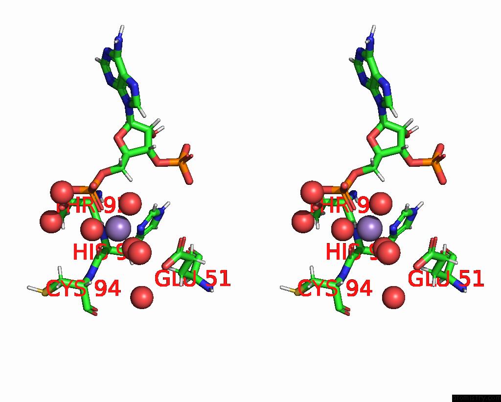
Manganese binding site 2 out of 8 in 8qzj
Go back to
Manganese binding site 2 out
of 8 in the Crystal Structure of Pptt-Adp From Mycobacterium Tuberculosis
Mono view
Stereo pair view
Mono view
Stereo pair view
A full contact list of Manganese with other atoms in the Mn binding
site number 2 of Crystal Structure of Pptt-Adp From Mycobacterium Tuberculosis within 5.0Å range:
|
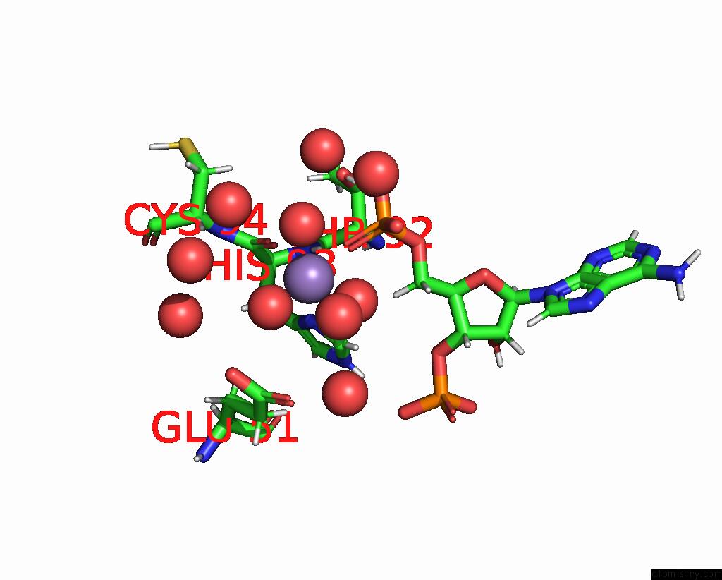
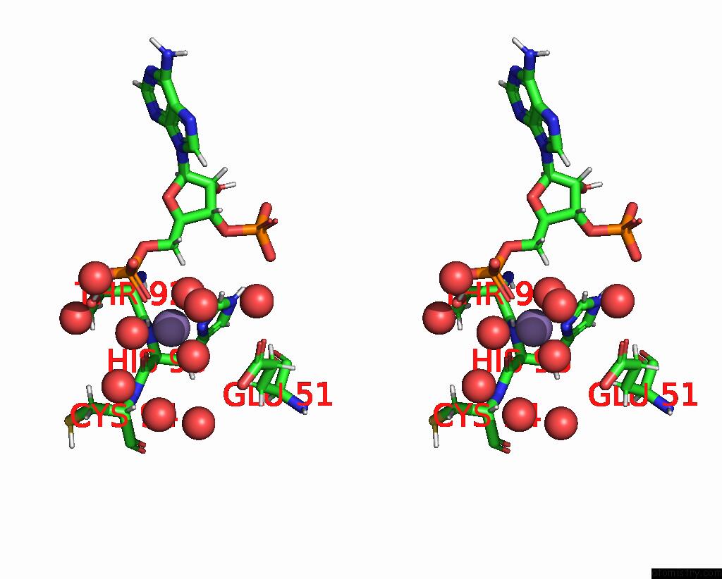
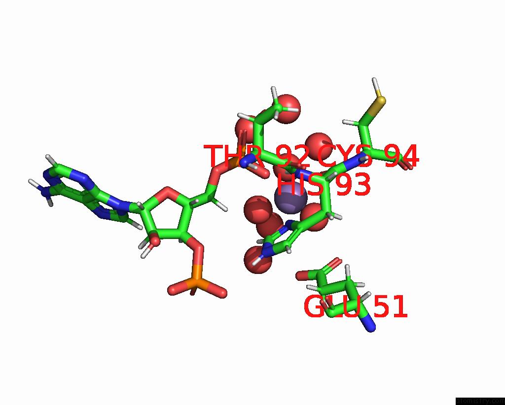
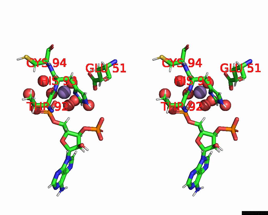
Manganese binding site 3 out of 8 in 8qzj
Go back to
Manganese binding site 3 out
of 8 in the Crystal Structure of Pptt-Adp From Mycobacterium Tuberculosis
Mono view
Stereo pair view
Mono view
Stereo pair view
A full contact list of Manganese with other atoms in the Mn binding
site number 3 of Crystal Structure of Pptt-Adp From Mycobacterium Tuberculosis within 5.0Å range:
|
Manganese binding site 4 out of 8 in 8qzj
Go back to
Manganese binding site 4 out
of 8 in the Crystal Structure of Pptt-Adp From Mycobacterium Tuberculosis
Mono view
Stereo pair view
Mono view
Stereo pair view
A full contact list of Manganese with other atoms in the Mn binding
site number 4 of Crystal Structure of Pptt-Adp From Mycobacterium Tuberculosis within 5.0Å range:
|
Manganese binding site 5 out of 8 in 8qzj
Go back to
Manganese binding site 5 out
of 8 in the Crystal Structure of Pptt-Adp From Mycobacterium Tuberculosis
Mono view
Stereo pair view
Mono view
Stereo pair view
A full contact list of Manganese with other atoms in the Mn binding
site number 5 of Crystal Structure of Pptt-Adp From Mycobacterium Tuberculosis within 5.0Å range:
|
Manganese binding site 6 out of 8 in 8qzj
Go back to
Manganese binding site 6 out
of 8 in the Crystal Structure of Pptt-Adp From Mycobacterium Tuberculosis
Mono view
Stereo pair view
Mono view
Stereo pair view
A full contact list of Manganese with other atoms in the Mn binding
site number 6 of Crystal Structure of Pptt-Adp From Mycobacterium Tuberculosis within 5.0Å range:
|
Manganese binding site 7 out of 8 in 8qzj
Go back to
Manganese binding site 7 out
of 8 in the Crystal Structure of Pptt-Adp From Mycobacterium Tuberculosis
Mono view
Stereo pair view
Mono view
Stereo pair view
A full contact list of Manganese with other atoms in the Mn binding
site number 7 of Crystal Structure of Pptt-Adp From Mycobacterium Tuberculosis within 5.0Å range:
|
Manganese binding site 8 out of 8 in 8qzj
Go back to
Manganese binding site 8 out
of 8 in the Crystal Structure of Pptt-Adp From Mycobacterium Tuberculosis
Mono view
Stereo pair view
Mono view
Stereo pair view
A full contact list of Manganese with other atoms in the Mn binding
site number 8 of Crystal Structure of Pptt-Adp From Mycobacterium Tuberculosis within 5.0Å range:
|
Reference:
S.Gavalda,
A.Faille,
S.Fioccola,
M.C.Nguyen,
C.Carivenc,
K.Rottier,
Y.Rufin,
S.Saitta,
G.Czaplicki,
C.Guilhot,
C.Chalut,
M.Brut,
L.Mourey,
J.D.Pedelacq.
Catalytic Cycle of Type II 4'-Phosphopantetheinyl Transferases Acs Catalysis V. 14 8561 2024.
ISSN: ESSN 2155-5435
DOI: 10.1021/ACSCATAL.3C06249
Page generated: Sun Oct 6 13:45:12 2024
ISSN: ESSN 2155-5435
DOI: 10.1021/ACSCATAL.3C06249
Last articles
Zn in 9MJ5Zn in 9HNW
Zn in 9G0L
Zn in 9FNE
Zn in 9DZN
Zn in 9E0I
Zn in 9D32
Zn in 9DAK
Zn in 8ZXC
Zn in 8ZUF