Manganese »
PDB 8q3z-8slm »
8q42 »
Manganese in PDB 8q42: Crystal Structure of CA4-Bound CAN2 (E341A) in Complex with Oligo-A Dna
Protein crystallography data
The structure of Crystal Structure of CA4-Bound CAN2 (E341A) in Complex with Oligo-A Dna, PDB code: 8q42
was solved by
K.Jungfer,
A.Sigg,
M.Jinek,
with X-Ray Crystallography technique. A brief refinement statistics is given in the table below:
| Resolution Low / High (Å) | 47.23 / 1.97 |
| Space group | P 1 21 1 |
| Cell size a, b, c (Å), α, β, γ (°) | 57.897, 78.026, 94.791, 90, 95.45, 90 |
| R / Rfree (%) | 18.6 / 21.7 |
Manganese Binding Sites:
The binding sites of Manganese atom in the Crystal Structure of CA4-Bound CAN2 (E341A) in Complex with Oligo-A Dna
(pdb code 8q42). This binding sites where shown within
5.0 Angstroms radius around Manganese atom.
In total 2 binding sites of Manganese where determined in the Crystal Structure of CA4-Bound CAN2 (E341A) in Complex with Oligo-A Dna, PDB code: 8q42:
Jump to Manganese binding site number: 1; 2;
In total 2 binding sites of Manganese where determined in the Crystal Structure of CA4-Bound CAN2 (E341A) in Complex with Oligo-A Dna, PDB code: 8q42:
Jump to Manganese binding site number: 1; 2;
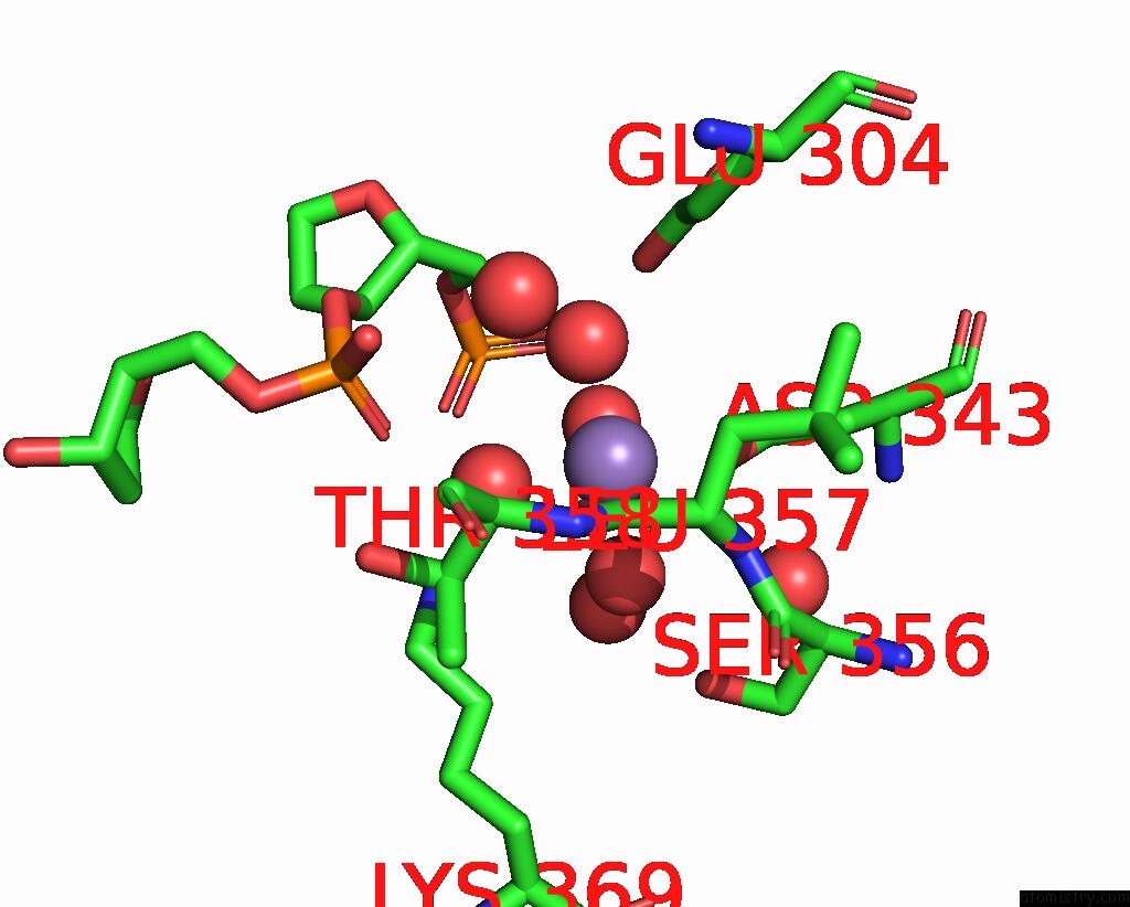
Manganese binding site 1 out of 2 in 8q42
Go back to
Manganese binding site 1 out
of 2 in the Crystal Structure of CA4-Bound CAN2 (E341A) in Complex with Oligo-A Dna
Mono view
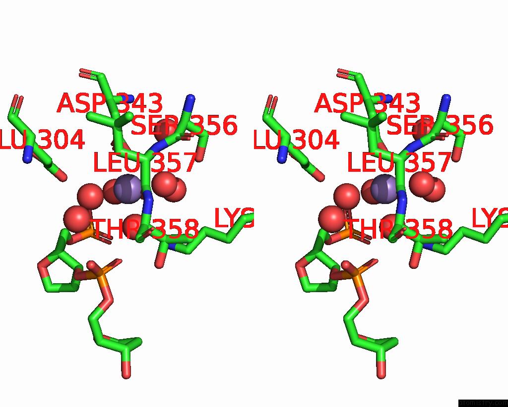
Stereo pair view
Mono view
Stereo pair view
A full contact list of Manganese with other atoms in the Mn binding
site number 1 of Crystal Structure of CA4-Bound CAN2 (E341A) in Complex with Oligo-A Dna within 5.0Å range:
|
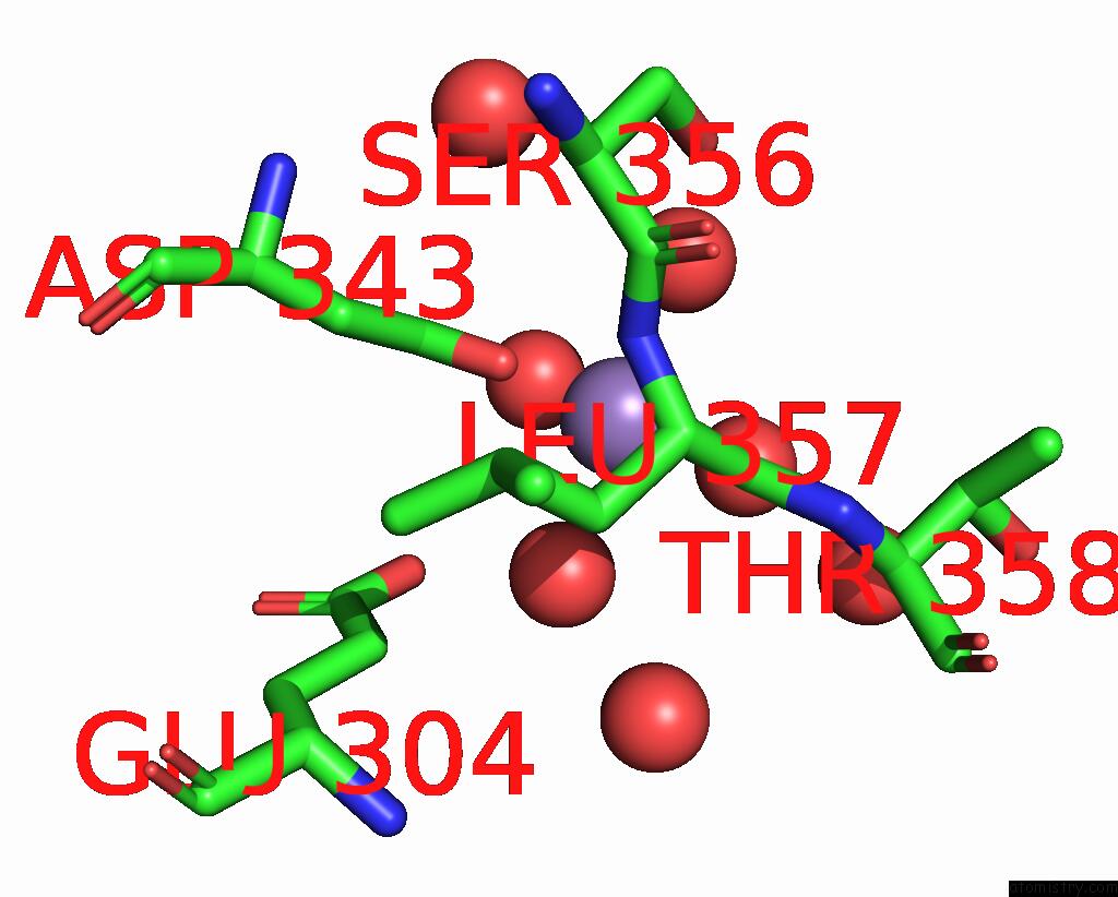
Manganese binding site 2 out of 2 in 8q42
Go back to
Manganese binding site 2 out
of 2 in the Crystal Structure of CA4-Bound CAN2 (E341A) in Complex with Oligo-A Dna
Mono view

Stereo pair view
Mono view
Stereo pair view
A full contact list of Manganese with other atoms in the Mn binding
site number 2 of Crystal Structure of CA4-Bound CAN2 (E341A) in Complex with Oligo-A Dna within 5.0Å range:
|
Reference:
K.Jungfer,
A.Sigg,
M.Jinek.
Substrate Selectivity and Catalytic Activation of the Type III Crispr-Associated Ancillary Nuclease CAN2 To Be Published.
Page generated: Sun Oct 6 13:38:47 2024
Last articles
F in 7L5EF in 7L72
F in 7L5P
F in 7L69
F in 7L5O
F in 7L0K
F in 7L4W
F in 7L4U
F in 7L4N
F in 7L4M