Manganese »
PDB 8kfu-8q1x »
8oz8 »
Manganese in PDB 8oz8: Crystal Structure of An Hydroxynitrile Lyase Variant (H96A) From Granulicella Tundricola
Protein crystallography data
The structure of Crystal Structure of An Hydroxynitrile Lyase Variant (H96A) From Granulicella Tundricola, PDB code: 8oz8
was solved by
I.Bento,
J.Coloma,
P.-L.Hagedoorn,
U.Hanefeld,
with X-Ray Crystallography technique. A brief refinement statistics is given in the table below:
| Resolution Low / High (Å) | 65.84 / 1.85 |
| Space group | P 65 2 2 |
| Cell size a, b, c (Å), α, β, γ (°) | 152.049, 152.049, 109.92, 90, 90, 120 |
| R / Rfree (%) | 16.4 / 19.5 |
Other elements in 8oz8:
The structure of Crystal Structure of An Hydroxynitrile Lyase Variant (H96A) From Granulicella Tundricola also contains other interesting chemical elements:
| Bromine | (Br) | 3 atoms |
Manganese Binding Sites:
The binding sites of Manganese atom in the Crystal Structure of An Hydroxynitrile Lyase Variant (H96A) From Granulicella Tundricola
(pdb code 8oz8). This binding sites where shown within
5.0 Angstroms radius around Manganese atom.
In total 4 binding sites of Manganese where determined in the Crystal Structure of An Hydroxynitrile Lyase Variant (H96A) From Granulicella Tundricola, PDB code: 8oz8:
Jump to Manganese binding site number: 1; 2; 3; 4;
In total 4 binding sites of Manganese where determined in the Crystal Structure of An Hydroxynitrile Lyase Variant (H96A) From Granulicella Tundricola, PDB code: 8oz8:
Jump to Manganese binding site number: 1; 2; 3; 4;
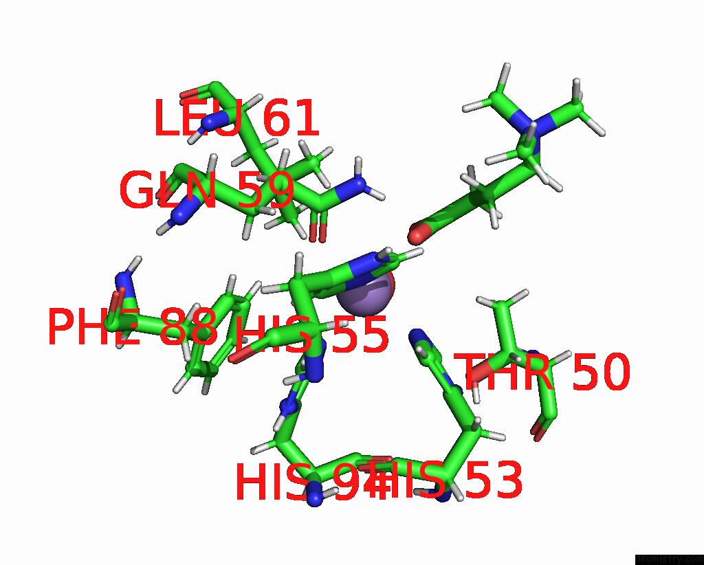
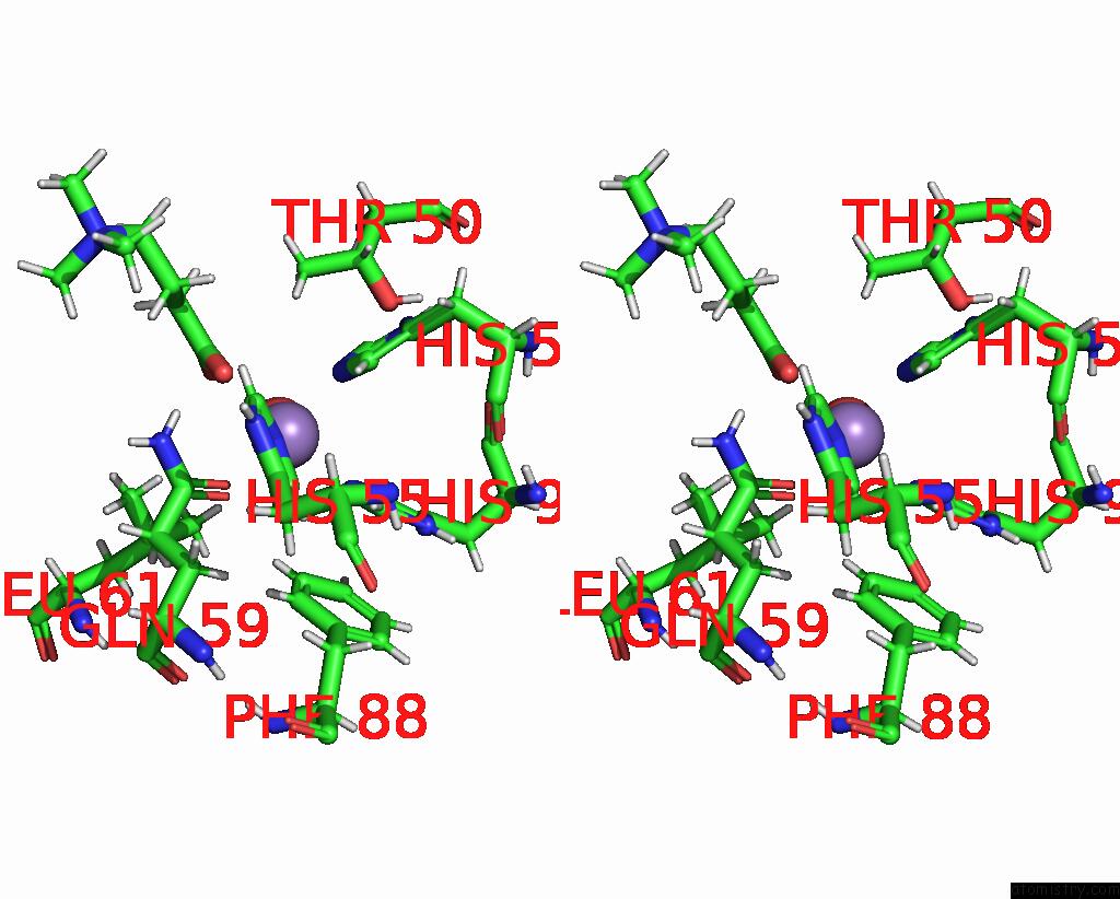
Manganese binding site 1 out of 4 in 8oz8
Go back to
Manganese binding site 1 out
of 4 in the Crystal Structure of An Hydroxynitrile Lyase Variant (H96A) From Granulicella Tundricola
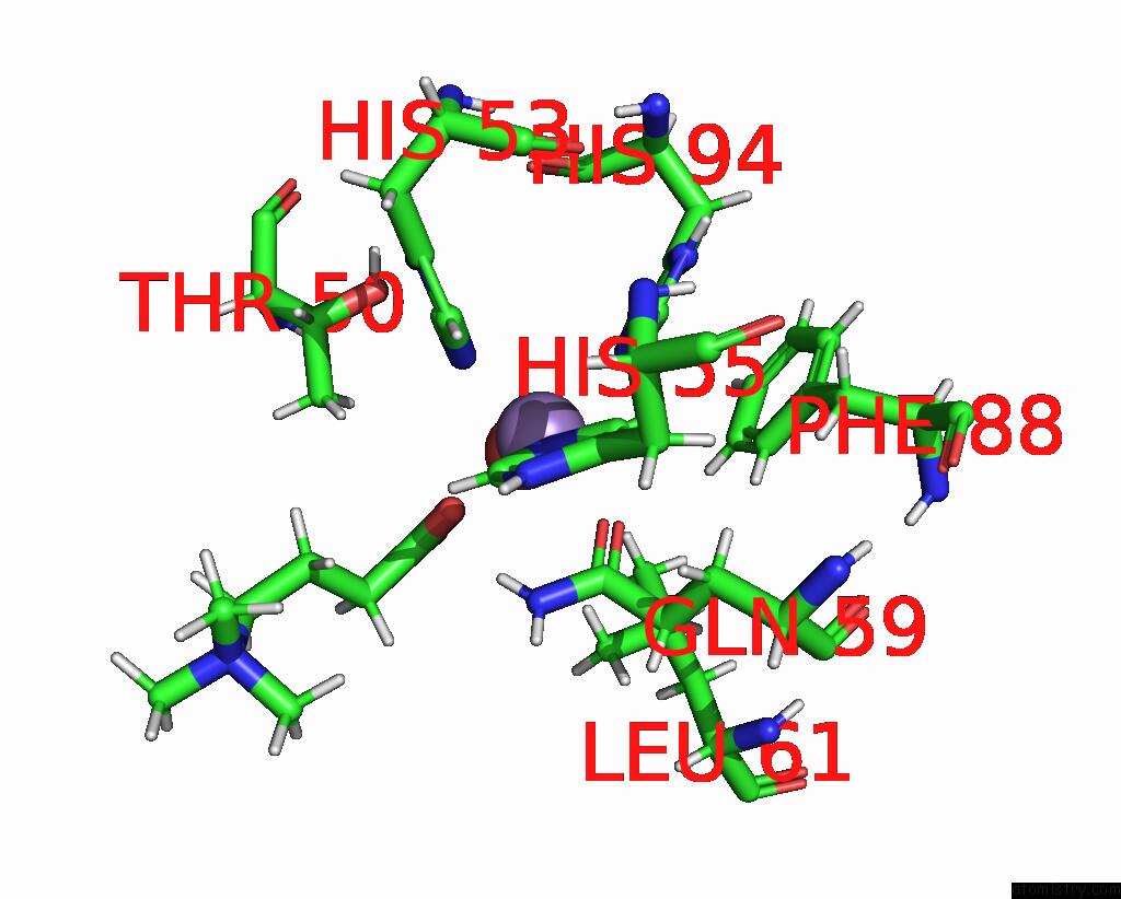
Mono view
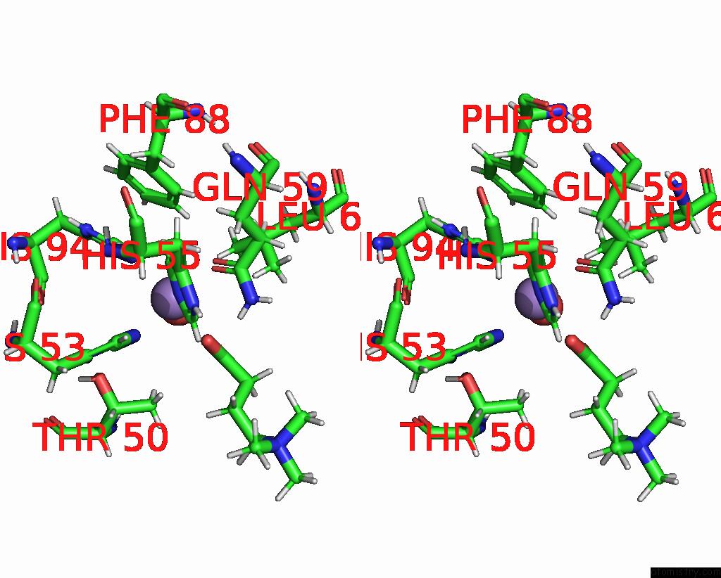
Stereo pair view
Mono view
Stereo pair view
A full contact list of Manganese with other atoms in the Mn binding
site number 1 of Crystal Structure of An Hydroxynitrile Lyase Variant (H96A) From Granulicella Tundricola within 5.0Å range:
|
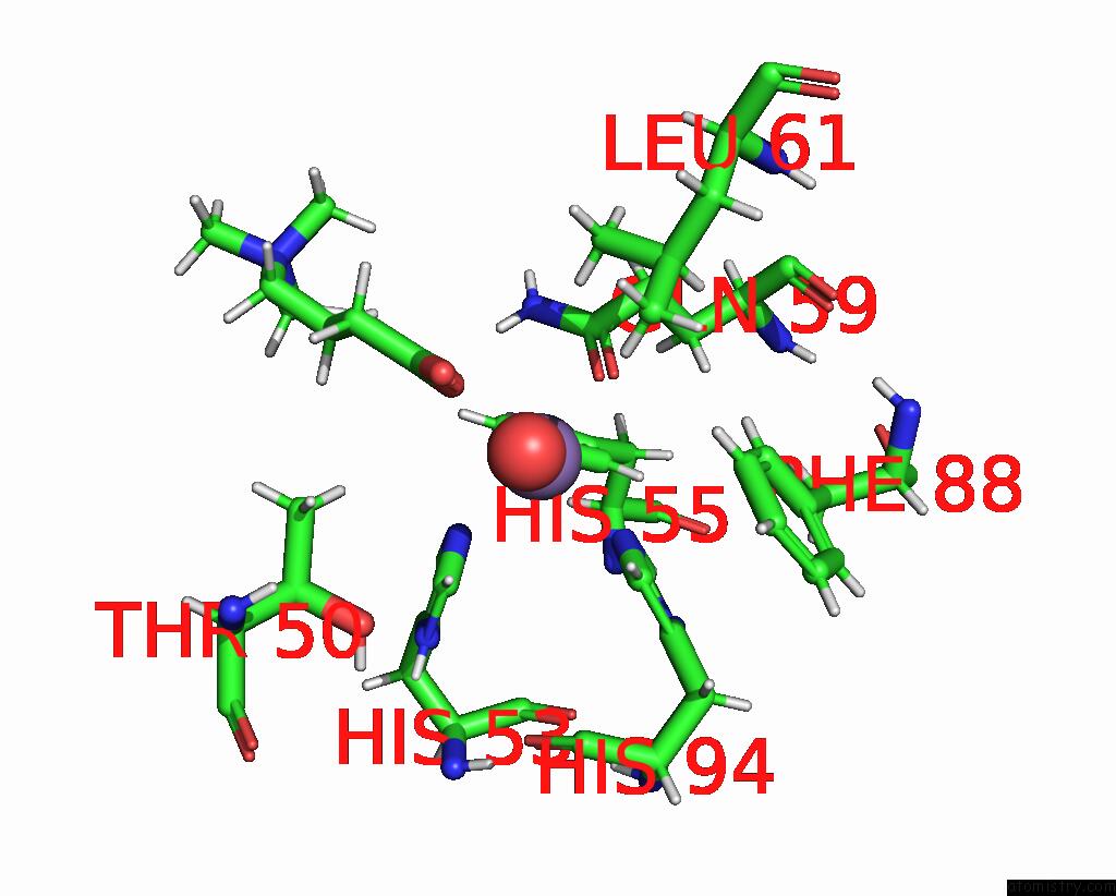
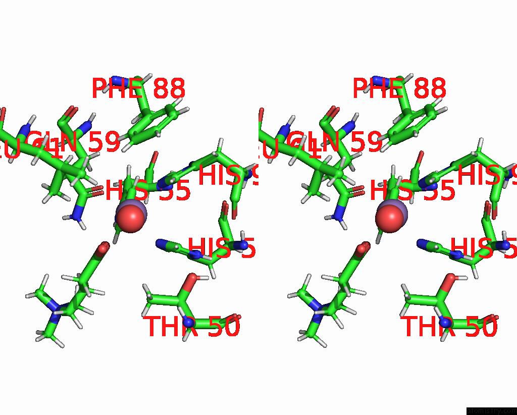
Manganese binding site 2 out of 4 in 8oz8
Go back to
Manganese binding site 2 out
of 4 in the Crystal Structure of An Hydroxynitrile Lyase Variant (H96A) From Granulicella Tundricola
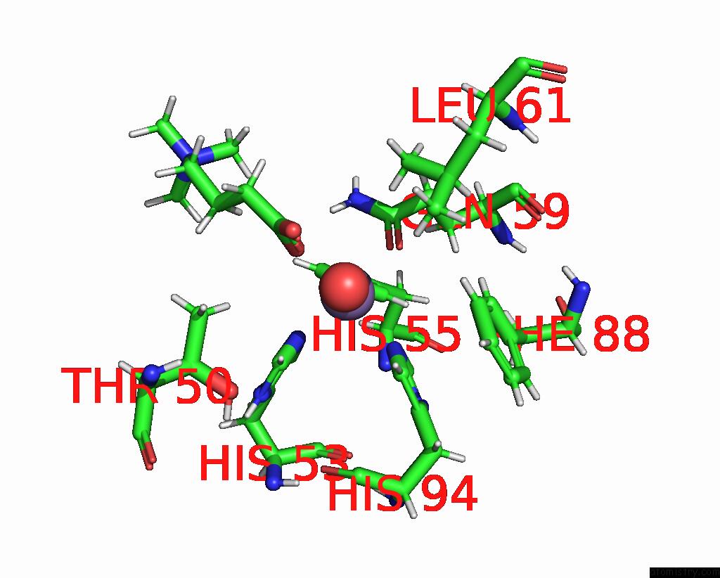
Mono view
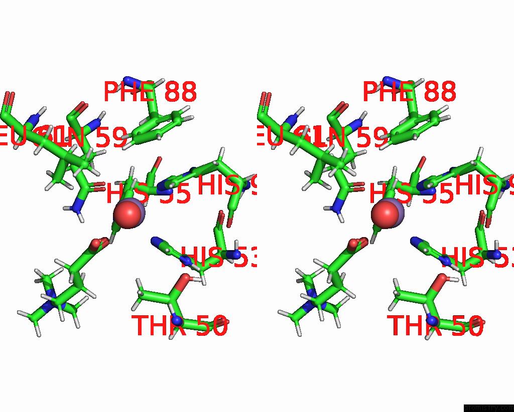
Stereo pair view
Mono view
Stereo pair view
A full contact list of Manganese with other atoms in the Mn binding
site number 2 of Crystal Structure of An Hydroxynitrile Lyase Variant (H96A) From Granulicella Tundricola within 5.0Å range:
|
Manganese binding site 3 out of 4 in 8oz8
Go back to
Manganese binding site 3 out
of 4 in the Crystal Structure of An Hydroxynitrile Lyase Variant (H96A) From Granulicella Tundricola
Mono view
Stereo pair view
Mono view
Stereo pair view
A full contact list of Manganese with other atoms in the Mn binding
site number 3 of Crystal Structure of An Hydroxynitrile Lyase Variant (H96A) From Granulicella Tundricola within 5.0Å range:
|
Manganese binding site 4 out of 4 in 8oz8
Go back to
Manganese binding site 4 out
of 4 in the Crystal Structure of An Hydroxynitrile Lyase Variant (H96A) From Granulicella Tundricola
Mono view
Stereo pair view
Mono view
Stereo pair view
A full contact list of Manganese with other atoms in the Mn binding
site number 4 of Crystal Structure of An Hydroxynitrile Lyase Variant (H96A) From Granulicella Tundricola within 5.0Å range:
|
Reference:
J.Coloma,
P.L.Hagedoorn,
I.Bento,
U.Hanefeld.
Can A Hydroxynitrile Lyase Catalyze An Oxidative Cleavage? Acs Catalysis V. 13 11182 2023.
ISSN: ESSN 2155-5435
DOI: 10.1021/ACSCATAL.3C02249
Page generated: Sun Oct 6 13:30:53 2024
ISSN: ESSN 2155-5435
DOI: 10.1021/ACSCATAL.3C02249
Last articles
Ca in 5S5XCa in 5S5Y
Ca in 5S5W
Ca in 5S5V
Ca in 5S5U
Ca in 5S5T
Ca in 5S5S
Ca in 5S5R
Ca in 5S5Q
Ca in 5S5P