Manganese »
PDB 8kfu-8q1x »
8oxg »
Manganese in PDB 8oxg: Crystal Structure of Human Methionine Aminopeptidase-2 Complexed with (3R,4S,5S,6R)-5-Methoxy-4-[(2R,3R)-2-Methyl-3-(3-Methyl-2-Buten-1- Yl)-2-Oxiranyl]-1-Oxaspiro[2.5]Oct-6-Yl N-(Trans-4-Aminocyclohexyl) Carbamate
Enzymatic activity of Crystal Structure of Human Methionine Aminopeptidase-2 Complexed with (3R,4S,5S,6R)-5-Methoxy-4-[(2R,3R)-2-Methyl-3-(3-Methyl-2-Buten-1- Yl)-2-Oxiranyl]-1-Oxaspiro[2.5]Oct-6-Yl N-(Trans-4-Aminocyclohexyl) Carbamate
All present enzymatic activity of Crystal Structure of Human Methionine Aminopeptidase-2 Complexed with (3R,4S,5S,6R)-5-Methoxy-4-[(2R,3R)-2-Methyl-3-(3-Methyl-2-Buten-1- Yl)-2-Oxiranyl]-1-Oxaspiro[2.5]Oct-6-Yl N-(Trans-4-Aminocyclohexyl) Carbamate:
3.4.11.18;
3.4.11.18;
Protein crystallography data
The structure of Crystal Structure of Human Methionine Aminopeptidase-2 Complexed with (3R,4S,5S,6R)-5-Methoxy-4-[(2R,3R)-2-Methyl-3-(3-Methyl-2-Buten-1- Yl)-2-Oxiranyl]-1-Oxaspiro[2.5]Oct-6-Yl N-(Trans-4-Aminocyclohexyl) Carbamate, PDB code: 8oxg
was solved by
S.Moss,
P.Cornelius,
with X-Ray Crystallography technique. A brief refinement statistics is given in the table below:
| Resolution Low / High (Å) | 44.84 / 1.73 |
| Space group | C 1 2 1 |
| Cell size a, b, c (Å), α, β, γ (°) | 119.05, 101.127, 83.737, 90, 98.76, 90 |
| R / Rfree (%) | 18.1 / 21.5 |
Manganese Binding Sites:
The binding sites of Manganese atom in the Crystal Structure of Human Methionine Aminopeptidase-2 Complexed with (3R,4S,5S,6R)-5-Methoxy-4-[(2R,3R)-2-Methyl-3-(3-Methyl-2-Buten-1- Yl)-2-Oxiranyl]-1-Oxaspiro[2.5]Oct-6-Yl N-(Trans-4-Aminocyclohexyl) Carbamate
(pdb code 8oxg). This binding sites where shown within
5.0 Angstroms radius around Manganese atom.
In total 4 binding sites of Manganese where determined in the Crystal Structure of Human Methionine Aminopeptidase-2 Complexed with (3R,4S,5S,6R)-5-Methoxy-4-[(2R,3R)-2-Methyl-3-(3-Methyl-2-Buten-1- Yl)-2-Oxiranyl]-1-Oxaspiro[2.5]Oct-6-Yl N-(Trans-4-Aminocyclohexyl) Carbamate, PDB code: 8oxg:
Jump to Manganese binding site number: 1; 2; 3; 4;
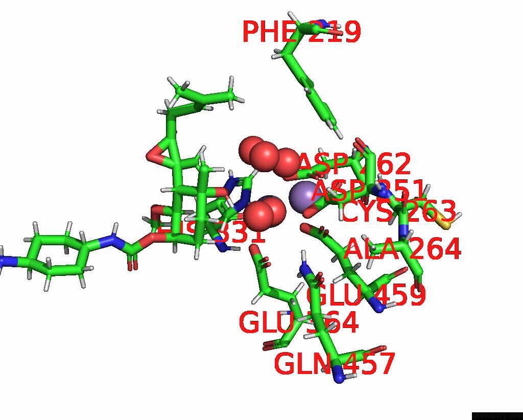
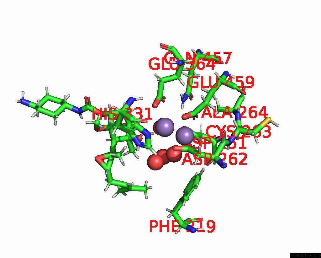
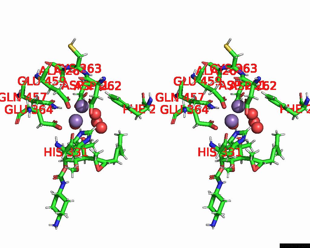
Manganese binding site 1 out of 4 in 8oxg
Go back to
Manganese binding site 1 out
of 4 in the Crystal Structure of Human Methionine Aminopeptidase-2 Complexed with (3R,4S,5S,6R)-5-Methoxy-4-[(2R,3R)-2-Methyl-3-(3-Methyl-2-Buten-1- Yl)-2-Oxiranyl]-1-Oxaspiro[2.5]Oct-6-Yl N-(Trans-4-Aminocyclohexyl) Carbamate
Mono view
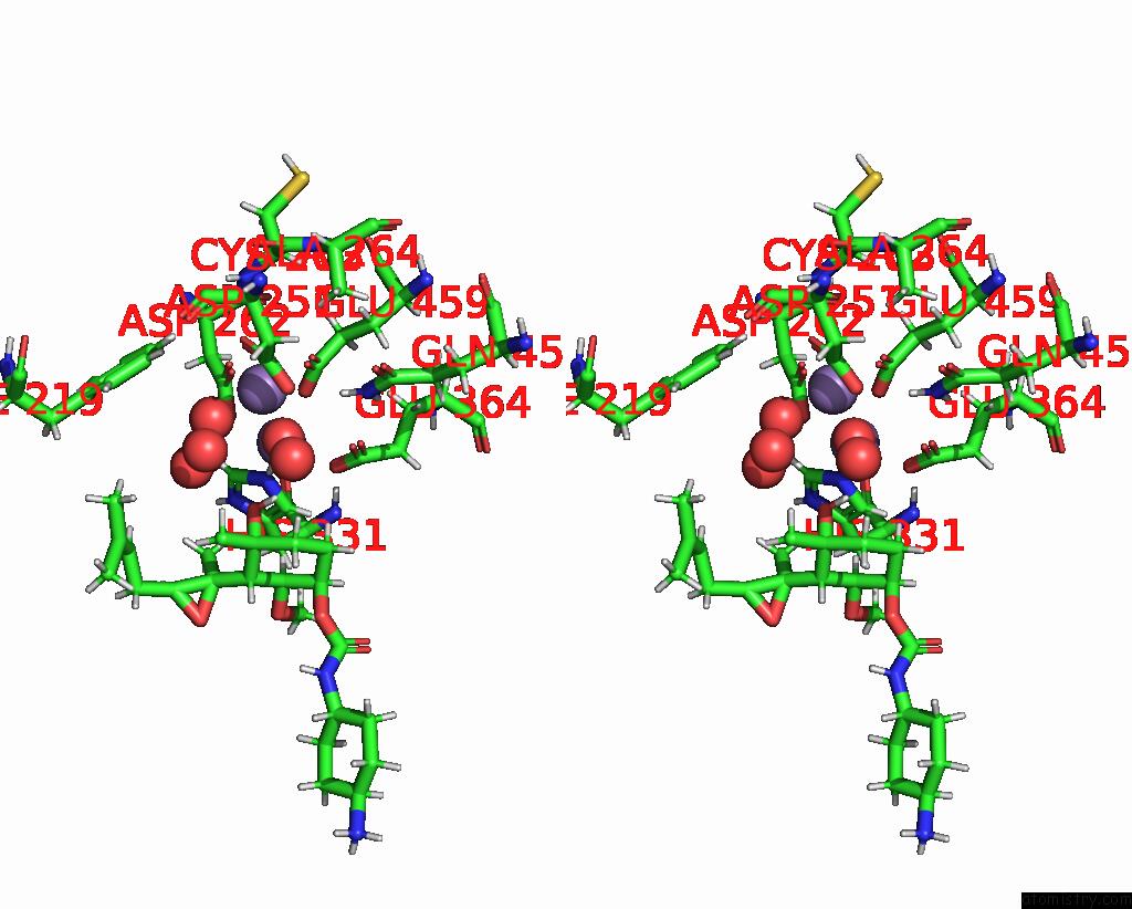
Stereo pair view
Mono view
Stereo pair view
A full contact list of Manganese with other atoms in the Mn binding
site number 1 of Crystal Structure of Human Methionine Aminopeptidase-2 Complexed with (3R,4S,5S,6R)-5-Methoxy-4-[(2R,3R)-2-Methyl-3-(3-Methyl-2-Buten-1- Yl)-2-Oxiranyl]-1-Oxaspiro[2.5]Oct-6-Yl N-(Trans-4-Aminocyclohexyl) Carbamate within 5.0Å range:
|
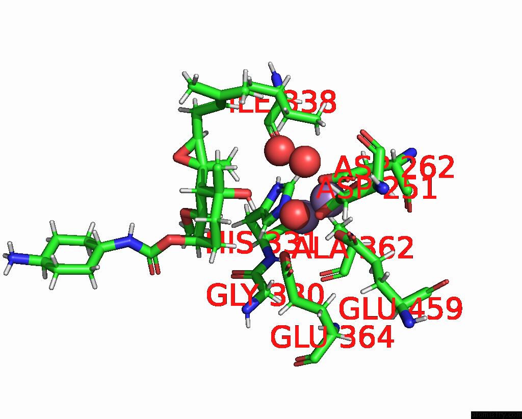
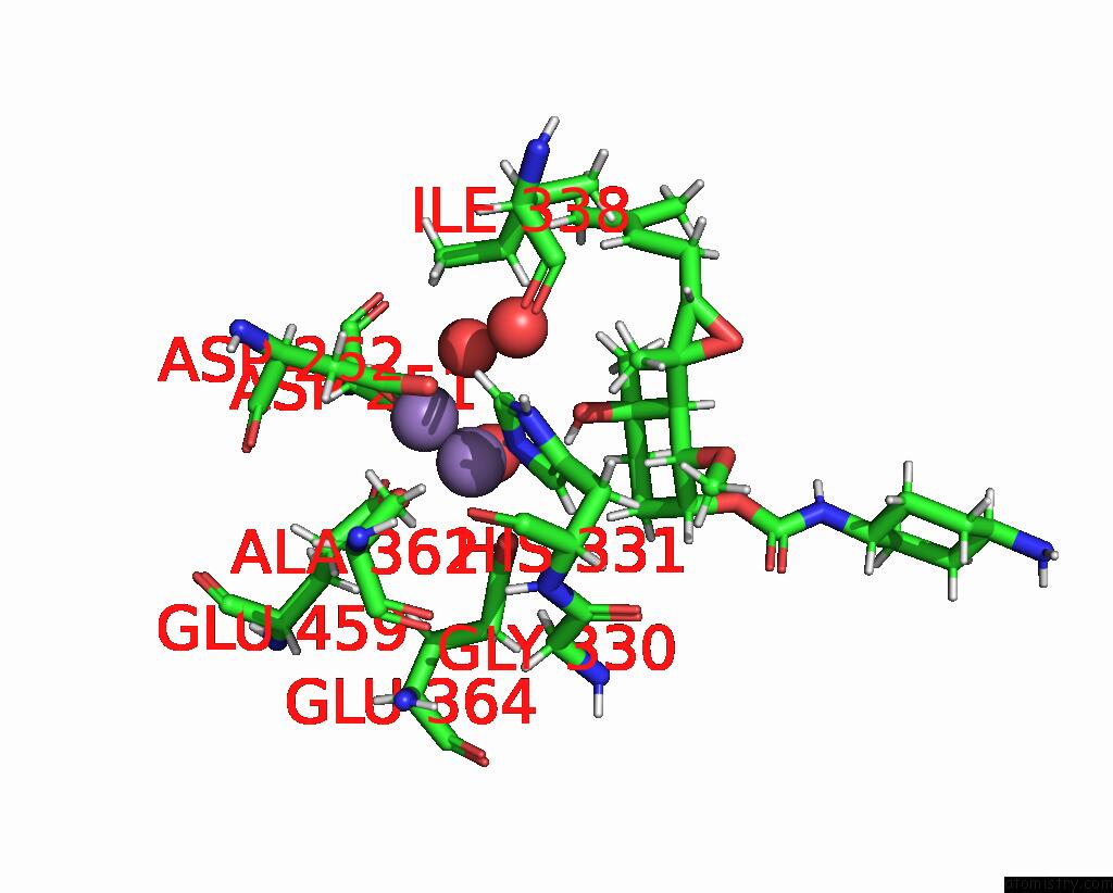
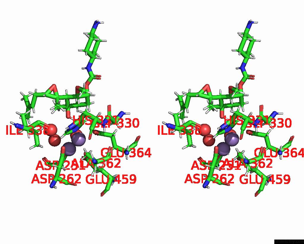
Manganese binding site 2 out of 4 in 8oxg
Go back to
Manganese binding site 2 out
of 4 in the Crystal Structure of Human Methionine Aminopeptidase-2 Complexed with (3R,4S,5S,6R)-5-Methoxy-4-[(2R,3R)-2-Methyl-3-(3-Methyl-2-Buten-1- Yl)-2-Oxiranyl]-1-Oxaspiro[2.5]Oct-6-Yl N-(Trans-4-Aminocyclohexyl) Carbamate
Mono view
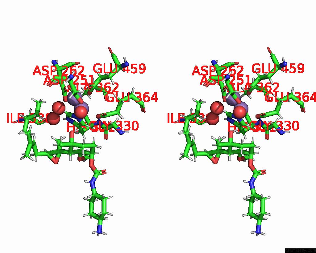
Stereo pair view
Mono view
Stereo pair view
A full contact list of Manganese with other atoms in the Mn binding
site number 2 of Crystal Structure of Human Methionine Aminopeptidase-2 Complexed with (3R,4S,5S,6R)-5-Methoxy-4-[(2R,3R)-2-Methyl-3-(3-Methyl-2-Buten-1- Yl)-2-Oxiranyl]-1-Oxaspiro[2.5]Oct-6-Yl N-(Trans-4-Aminocyclohexyl) Carbamate within 5.0Å range:
|
Manganese binding site 3 out of 4 in 8oxg
Go back to
Manganese binding site 3 out
of 4 in the Crystal Structure of Human Methionine Aminopeptidase-2 Complexed with (3R,4S,5S,6R)-5-Methoxy-4-[(2R,3R)-2-Methyl-3-(3-Methyl-2-Buten-1- Yl)-2-Oxiranyl]-1-Oxaspiro[2.5]Oct-6-Yl N-(Trans-4-Aminocyclohexyl) Carbamate
Mono view
Stereo pair view
Mono view
Stereo pair view
A full contact list of Manganese with other atoms in the Mn binding
site number 3 of Crystal Structure of Human Methionine Aminopeptidase-2 Complexed with (3R,4S,5S,6R)-5-Methoxy-4-[(2R,3R)-2-Methyl-3-(3-Methyl-2-Buten-1- Yl)-2-Oxiranyl]-1-Oxaspiro[2.5]Oct-6-Yl N-(Trans-4-Aminocyclohexyl) Carbamate within 5.0Å range:
|
Manganese binding site 4 out of 4 in 8oxg
Go back to
Manganese binding site 4 out
of 4 in the Crystal Structure of Human Methionine Aminopeptidase-2 Complexed with (3R,4S,5S,6R)-5-Methoxy-4-[(2R,3R)-2-Methyl-3-(3-Methyl-2-Buten-1- Yl)-2-Oxiranyl]-1-Oxaspiro[2.5]Oct-6-Yl N-(Trans-4-Aminocyclohexyl) Carbamate
Mono view
Stereo pair view
Mono view
Stereo pair view
A full contact list of Manganese with other atoms in the Mn binding
site number 4 of Crystal Structure of Human Methionine Aminopeptidase-2 Complexed with (3R,4S,5S,6R)-5-Methoxy-4-[(2R,3R)-2-Methyl-3-(3-Methyl-2-Buten-1- Yl)-2-Oxiranyl]-1-Oxaspiro[2.5]Oct-6-Yl N-(Trans-4-Aminocyclohexyl) Carbamate within 5.0Å range:
|
Reference:
P.Cornelius,
B.A.Mayes,
J.S.Petersen,
D.J.Turnquist,
P.J.Dufour,
A.J.Dannenberg,
J.M.Shanahan,
B.J.Carver.
Pharmacological Characterization of Sdx-7320/Evexomostat: A Novel Methionine Aminopeptidase Type 2 Inhibitor with Anti-Tumor and Anti-Metastatic Activity. Mol.Cancer Ther. 2024.
ISSN: ESSN 1538-8514
PubMed: 38530115
DOI: 10.1158/1535-7163.MCT-23-0574
Page generated: Sun Oct 6 13:30:51 2024
ISSN: ESSN 1538-8514
PubMed: 38530115
DOI: 10.1158/1535-7163.MCT-23-0574
Last articles
Zn in 9J0NZn in 9J0O
Zn in 9J0P
Zn in 9FJX
Zn in 9EKB
Zn in 9C0F
Zn in 9CAH
Zn in 9CH0
Zn in 9CH3
Zn in 9CH1