Manganese »
PDB 8iri-8kft »
8jqe »
Manganese in PDB 8jqe: Structure of Cmcbda in Complex with MN2+ and Glycerol
Protein crystallography data
The structure of Structure of Cmcbda in Complex with MN2+ and Glycerol, PDB code: 8jqe
was solved by
X.Li,
with X-Ray Crystallography technique. A brief refinement statistics is given in the table below:
| Resolution Low / High (Å) | 34.99 / 2.31 |
| Space group | P 1 21 1 |
| Cell size a, b, c (Å), α, β, γ (°) | 67.617, 60.27, 87.444, 90, 111.11, 90 |
| R / Rfree (%) | 16 / 21.4 |
Manganese Binding Sites:
The binding sites of Manganese atom in the Structure of Cmcbda in Complex with MN2+ and Glycerol
(pdb code 8jqe). This binding sites where shown within
5.0 Angstroms radius around Manganese atom.
In total 2 binding sites of Manganese where determined in the Structure of Cmcbda in Complex with MN2+ and Glycerol, PDB code: 8jqe:
Jump to Manganese binding site number: 1; 2;
In total 2 binding sites of Manganese where determined in the Structure of Cmcbda in Complex with MN2+ and Glycerol, PDB code: 8jqe:
Jump to Manganese binding site number: 1; 2;
Manganese binding site 1 out of 2 in 8jqe
Go back to
Manganese binding site 1 out
of 2 in the Structure of Cmcbda in Complex with MN2+ and Glycerol
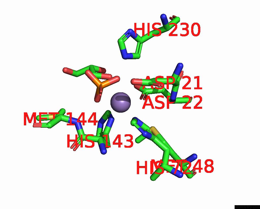
Mono view
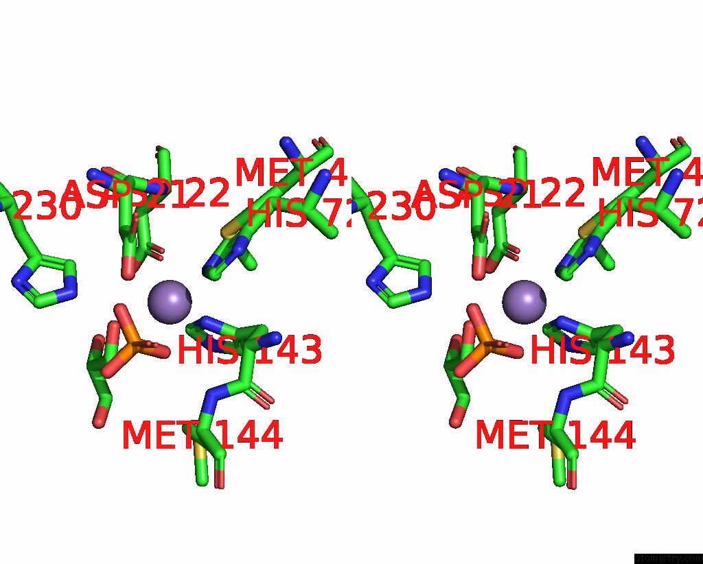
Stereo pair view
Mono view
Stereo pair view
A full contact list of Manganese with other atoms in the Mn binding
site number 1 of Structure of Cmcbda in Complex with MN2+ and Glycerol within 5.0Å range:
|
Manganese binding site 2 out of 2 in 8jqe
Go back to
Manganese binding site 2 out
of 2 in the Structure of Cmcbda in Complex with MN2+ and Glycerol

Mono view
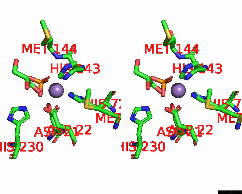
Stereo pair view
Mono view
Stereo pair view
A full contact list of Manganese with other atoms in the Mn binding
site number 2 of Structure of Cmcbda in Complex with MN2+ and Glycerol within 5.0Å range:
|
Reference:
S.Hu,
L.Xu,
C.Xie,
J.Hong.
Structural Insights Into the Catalytic Activity of Cyclobacterium Marinum N -Acetylglucosamine Deacetylase. J.Agric.Food Chem. 2023.
ISSN: ESSN 1520-5118
PubMed: 38141024
DOI: 10.1021/ACS.JAFC.3C06146
Page generated: Sun Oct 6 13:20:48 2024
ISSN: ESSN 1520-5118
PubMed: 38141024
DOI: 10.1021/ACS.JAFC.3C06146
Last articles
Zn in 9MJ5Zn in 9HNW
Zn in 9G0L
Zn in 9FNE
Zn in 9DZN
Zn in 9E0I
Zn in 9D32
Zn in 9DAK
Zn in 8ZXC
Zn in 8ZUF