Manganese »
PDB 8hlf-8irh »
8igi »
Manganese in PDB 8igi: Crystal Structure of HP1526 (Xtha)- A Base Excision Dna Repair Protein in Helicobacter Pylori
Protein crystallography data
The structure of Crystal Structure of HP1526 (Xtha)- A Base Excision Dna Repair Protein in Helicobacter Pylori, PDB code: 8igi
was solved by
T.T.Dinh,
O.Dao,
K.H.Lee,
with X-Ray Crystallography technique. A brief refinement statistics is given in the table below:
| Resolution Low / High (Å) | 37.05 / 1.84 |
| Space group | P 21 21 21 |
| Cell size a, b, c (Å), α, β, γ (°) | 72.096, 71.909, 108.033, 90, 90, 90 |
| R / Rfree (%) | 16.7 / 19.6 |
Manganese Binding Sites:
The binding sites of Manganese atom in the Crystal Structure of HP1526 (Xtha)- A Base Excision Dna Repair Protein in Helicobacter Pylori
(pdb code 8igi). This binding sites where shown within
5.0 Angstroms radius around Manganese atom.
In total 2 binding sites of Manganese where determined in the Crystal Structure of HP1526 (Xtha)- A Base Excision Dna Repair Protein in Helicobacter Pylori, PDB code: 8igi:
Jump to Manganese binding site number: 1; 2;
In total 2 binding sites of Manganese where determined in the Crystal Structure of HP1526 (Xtha)- A Base Excision Dna Repair Protein in Helicobacter Pylori, PDB code: 8igi:
Jump to Manganese binding site number: 1; 2;
Manganese binding site 1 out of 2 in 8igi
Go back to
Manganese binding site 1 out
of 2 in the Crystal Structure of HP1526 (Xtha)- A Base Excision Dna Repair Protein in Helicobacter Pylori
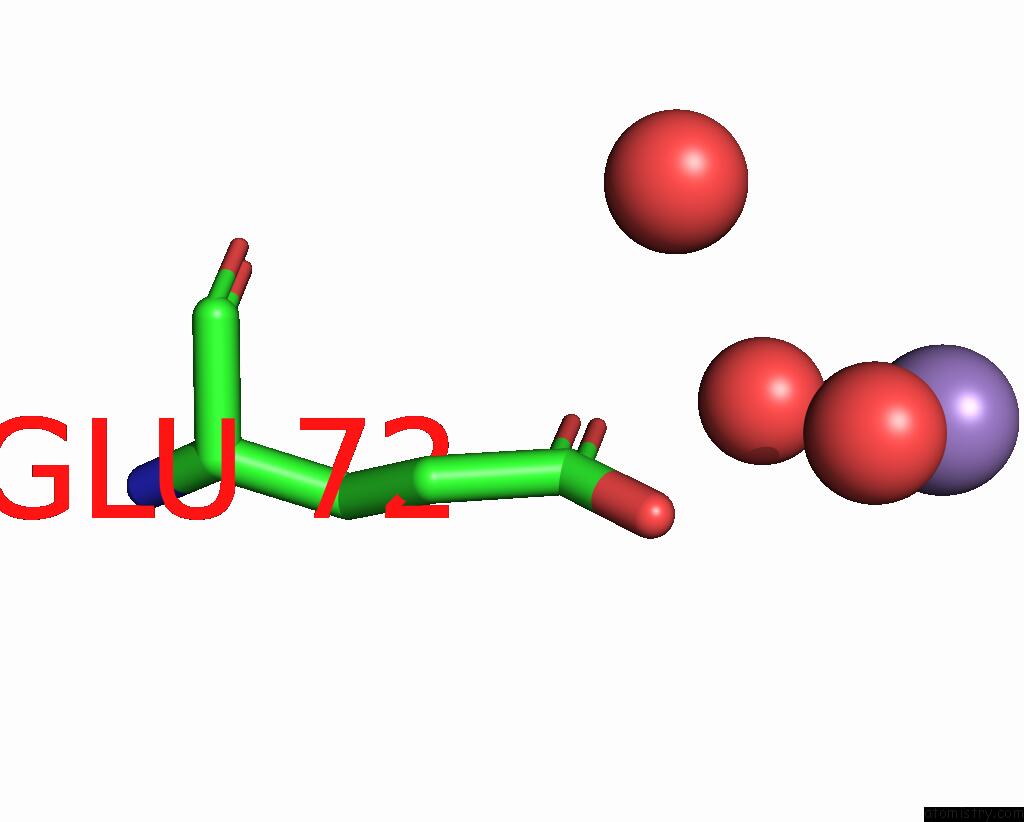
Mono view
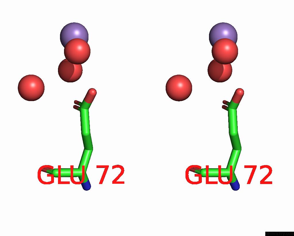
Stereo pair view
Mono view
Stereo pair view
A full contact list of Manganese with other atoms in the Mn binding
site number 1 of Crystal Structure of HP1526 (Xtha)- A Base Excision Dna Repair Protein in Helicobacter Pylori within 5.0Å range:
|
Manganese binding site 2 out of 2 in 8igi
Go back to
Manganese binding site 2 out
of 2 in the Crystal Structure of HP1526 (Xtha)- A Base Excision Dna Repair Protein in Helicobacter Pylori
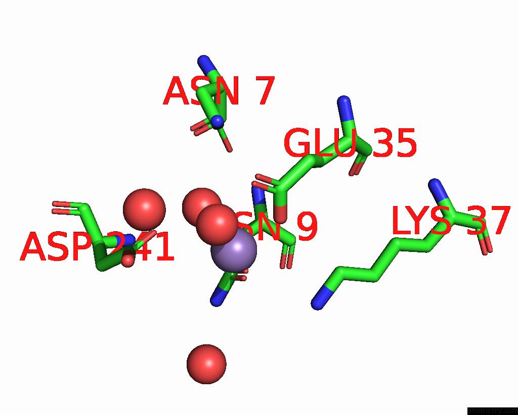
Mono view
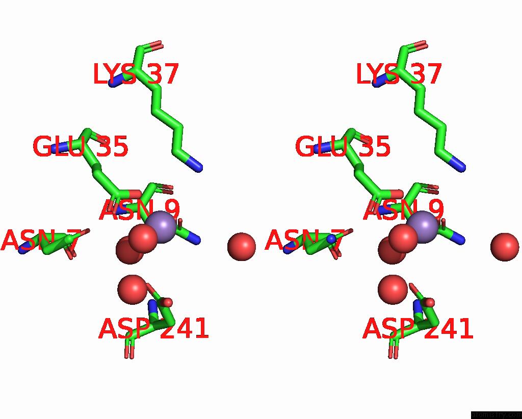
Stereo pair view
Mono view
Stereo pair view
A full contact list of Manganese with other atoms in the Mn binding
site number 2 of Crystal Structure of HP1526 (Xtha)- A Base Excision Dna Repair Protein in Helicobacter Pylori within 5.0Å range:
|
Reference:
T.Dinh,
O.Dao,
A.Killivalavan,
D.Ngo,
K.H.Lee.
Crystal Structure of the Apurinic/Apyrimidinic Endonuclease Xtha (HP1526 Protein) From Helicobacter Pylori. Biochem.Biophys.Res.Commun. V. 663 8 2023.
ISSN: ESSN 1090-2104
PubMed: 37116395
DOI: 10.1016/J.BBRC.2023.04.047
Page generated: Sun Oct 6 12:33:06 2024
ISSN: ESSN 1090-2104
PubMed: 37116395
DOI: 10.1016/J.BBRC.2023.04.047
Last articles
Zn in 9MJ5Zn in 9HNW
Zn in 9G0L
Zn in 9FNE
Zn in 9DZN
Zn in 9E0I
Zn in 9D32
Zn in 9DAK
Zn in 8ZXC
Zn in 8ZUF