Manganese »
PDB 8hlf-8irh »
8hmo »
Manganese in PDB 8hmo: Crystal Structure of Metal-Dependent Hydrolase Complexed with Manganese From Bacillus Smithii
Enzymatic activity of Crystal Structure of Metal-Dependent Hydrolase Complexed with Manganese From Bacillus Smithii
All present enzymatic activity of Crystal Structure of Metal-Dependent Hydrolase Complexed with Manganese From Bacillus Smithii:
3.5.1.9;
3.5.1.9;
Protein crystallography data
The structure of Crystal Structure of Metal-Dependent Hydrolase Complexed with Manganese From Bacillus Smithii, PDB code: 8hmo
was solved by
K.Yoneda,
H.Sakuraba,
T.Ohshima,
with X-Ray Crystallography technique. A brief refinement statistics is given in the table below:
| Resolution Low / High (Å) | 43.45 / 2.53 |
| Space group | P 1 2 1 |
| Cell size a, b, c (Å), α, β, γ (°) | 90.21, 70.198, 104.839, 90, 105.77, 90 |
| R / Rfree (%) | 25 / 29.9 |
Manganese Binding Sites:
Pages:
>>> Page 1 <<< Page 2, Binding sites: 11 - 12;Binding sites:
The binding sites of Manganese atom in the Crystal Structure of Metal-Dependent Hydrolase Complexed with Manganese From Bacillus Smithii (pdb code 8hmo). This binding sites where shown within 5.0 Angstroms radius around Manganese atom.In total 12 binding sites of Manganese where determined in the Crystal Structure of Metal-Dependent Hydrolase Complexed with Manganese From Bacillus Smithii, PDB code: 8hmo:
Jump to Manganese binding site number: 1; 2; 3; 4; 5; 6; 7; 8; 9; 10;
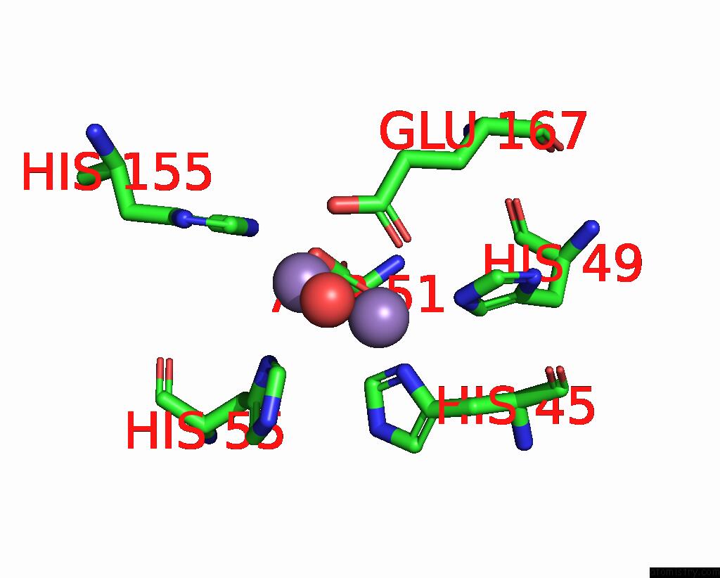
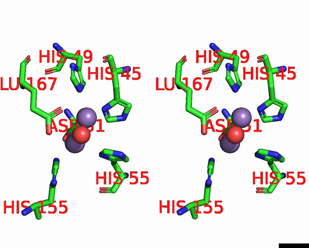
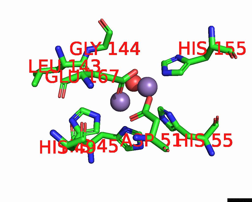
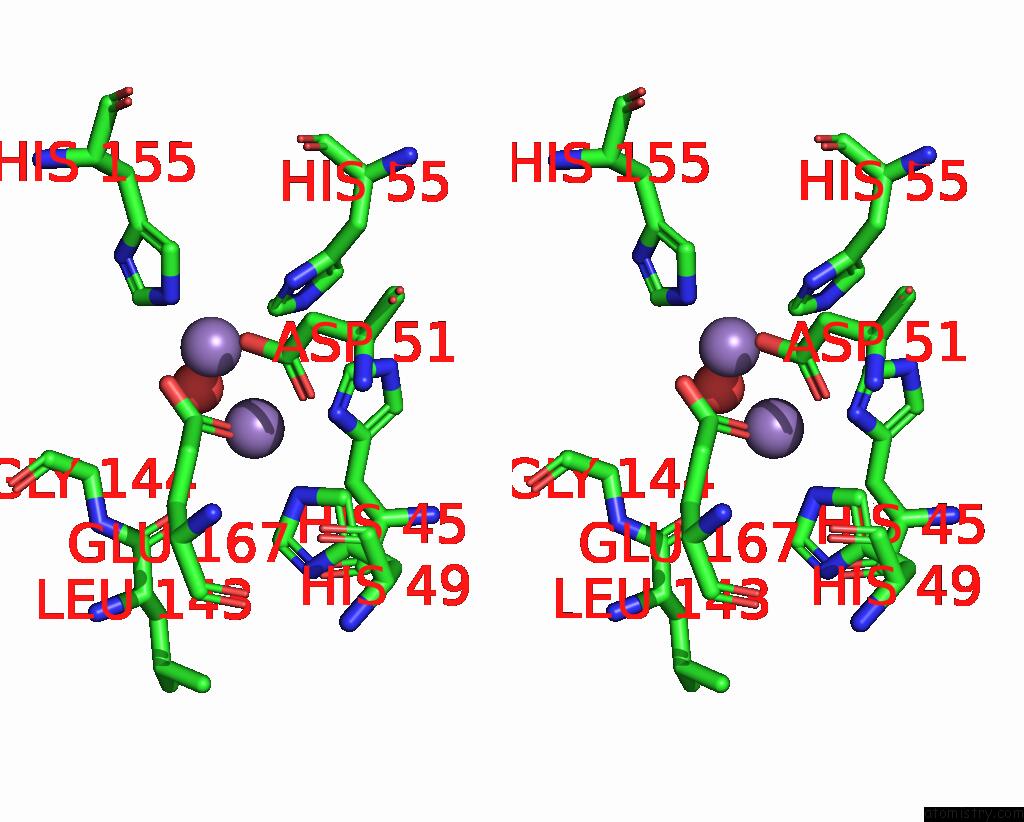
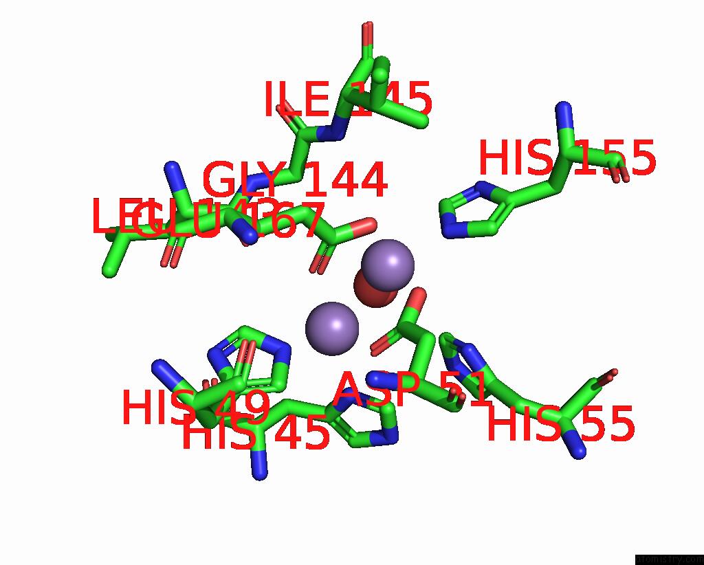
Manganese binding site 1 out of 12 in 8hmo
Go back to
Manganese binding site 1 out
of 12 in the Crystal Structure of Metal-Dependent Hydrolase Complexed with Manganese From Bacillus Smithii

Mono view
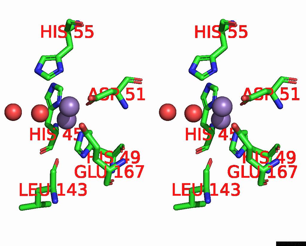
Stereo pair view
Mono view
Stereo pair view
A full contact list of Manganese with other atoms in the Mn binding
site number 1 of Crystal Structure of Metal-Dependent Hydrolase Complexed with Manganese From Bacillus Smithii within 5.0Å range:
|
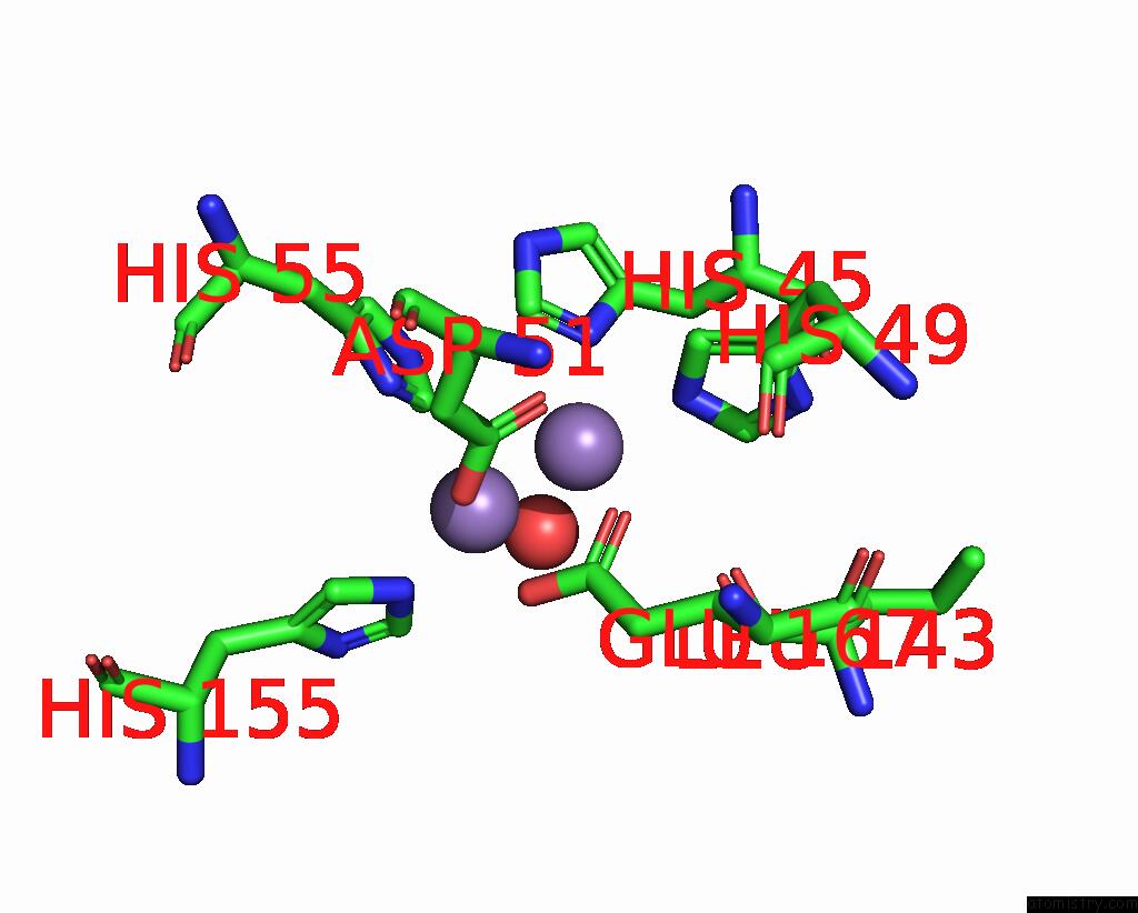
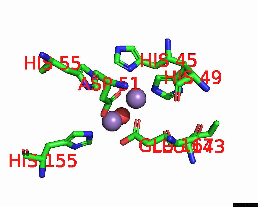
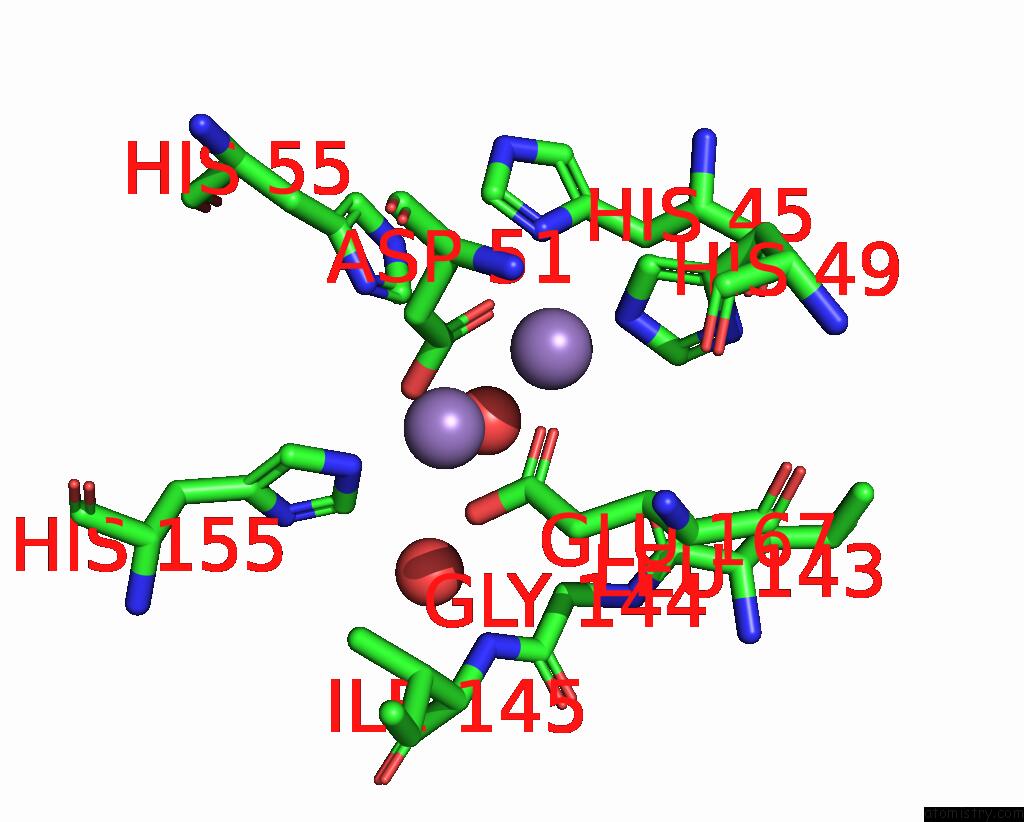
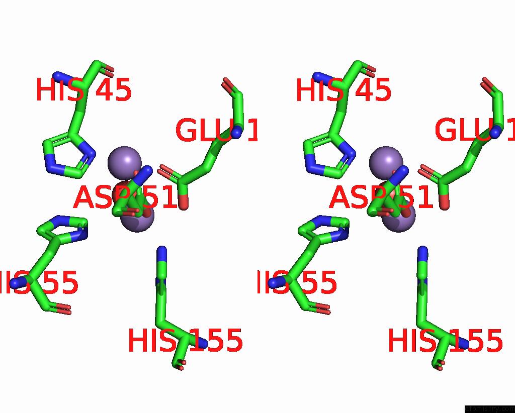
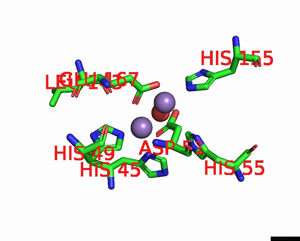
Manganese binding site 2 out of 12 in 8hmo
Go back to
Manganese binding site 2 out
of 12 in the Crystal Structure of Metal-Dependent Hydrolase Complexed with Manganese From Bacillus Smithii

Mono view
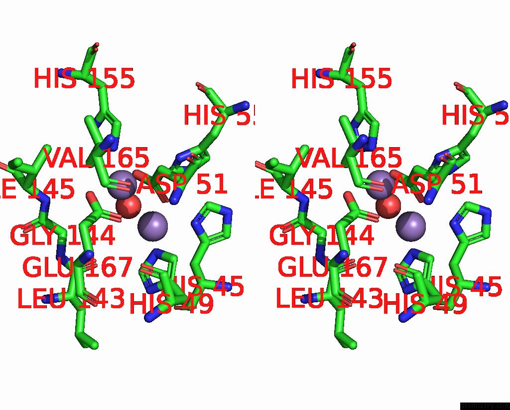
Stereo pair view
Mono view
Stereo pair view
A full contact list of Manganese with other atoms in the Mn binding
site number 2 of Crystal Structure of Metal-Dependent Hydrolase Complexed with Manganese From Bacillus Smithii within 5.0Å range:
|
Manganese binding site 3 out of 12 in 8hmo
Go back to
Manganese binding site 3 out
of 12 in the Crystal Structure of Metal-Dependent Hydrolase Complexed with Manganese From Bacillus Smithii
Mono view
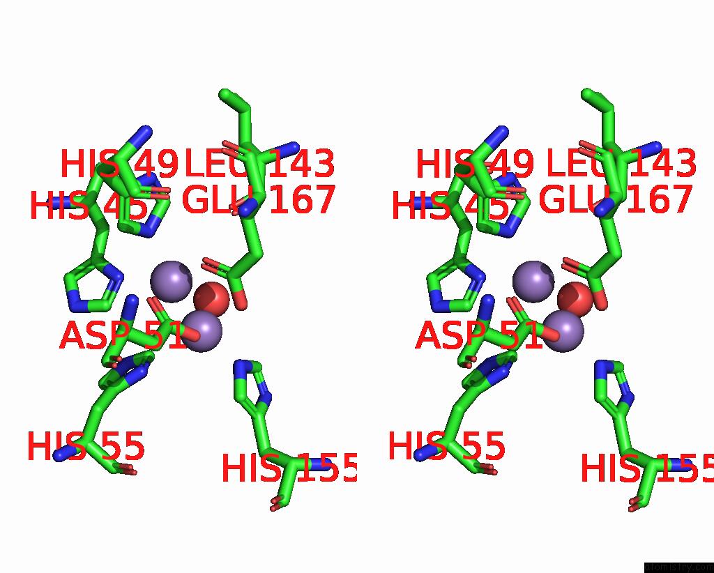
Stereo pair view
Mono view
Stereo pair view
A full contact list of Manganese with other atoms in the Mn binding
site number 3 of Crystal Structure of Metal-Dependent Hydrolase Complexed with Manganese From Bacillus Smithii within 5.0Å range:
|
Manganese binding site 4 out of 12 in 8hmo
Go back to
Manganese binding site 4 out
of 12 in the Crystal Structure of Metal-Dependent Hydrolase Complexed with Manganese From Bacillus Smithii
Mono view
Stereo pair view
Mono view
Stereo pair view
A full contact list of Manganese with other atoms in the Mn binding
site number 4 of Crystal Structure of Metal-Dependent Hydrolase Complexed with Manganese From Bacillus Smithii within 5.0Å range:
|
Manganese binding site 5 out of 12 in 8hmo
Go back to
Manganese binding site 5 out
of 12 in the Crystal Structure of Metal-Dependent Hydrolase Complexed with Manganese From Bacillus Smithii
Mono view

Stereo pair view
Mono view
Stereo pair view
A full contact list of Manganese with other atoms in the Mn binding
site number 5 of Crystal Structure of Metal-Dependent Hydrolase Complexed with Manganese From Bacillus Smithii within 5.0Å range:
|
Manganese binding site 6 out of 12 in 8hmo
Go back to
Manganese binding site 6 out
of 12 in the Crystal Structure of Metal-Dependent Hydrolase Complexed with Manganese From Bacillus Smithii
Mono view

Stereo pair view
Mono view
Stereo pair view
A full contact list of Manganese with other atoms in the Mn binding
site number 6 of Crystal Structure of Metal-Dependent Hydrolase Complexed with Manganese From Bacillus Smithii within 5.0Å range:
|
Manganese binding site 7 out of 12 in 8hmo
Go back to
Manganese binding site 7 out
of 12 in the Crystal Structure of Metal-Dependent Hydrolase Complexed with Manganese From Bacillus Smithii
Mono view
Stereo pair view
Mono view
Stereo pair view
A full contact list of Manganese with other atoms in the Mn binding
site number 7 of Crystal Structure of Metal-Dependent Hydrolase Complexed with Manganese From Bacillus Smithii within 5.0Å range:
|
Manganese binding site 8 out of 12 in 8hmo
Go back to
Manganese binding site 8 out
of 12 in the Crystal Structure of Metal-Dependent Hydrolase Complexed with Manganese From Bacillus Smithii

Mono view
Stereo pair view
Mono view
Stereo pair view
A full contact list of Manganese with other atoms in the Mn binding
site number 8 of Crystal Structure of Metal-Dependent Hydrolase Complexed with Manganese From Bacillus Smithii within 5.0Å range:
|
Manganese binding site 9 out of 12 in 8hmo
Go back to
Manganese binding site 9 out
of 12 in the Crystal Structure of Metal-Dependent Hydrolase Complexed with Manganese From Bacillus Smithii
Mono view

Stereo pair view
Mono view
Stereo pair view
A full contact list of Manganese with other atoms in the Mn binding
site number 9 of Crystal Structure of Metal-Dependent Hydrolase Complexed with Manganese From Bacillus Smithii within 5.0Å range:
|
Manganese binding site 10 out of 12 in 8hmo
Go back to
Manganese binding site 10 out
of 12 in the Crystal Structure of Metal-Dependent Hydrolase Complexed with Manganese From Bacillus Smithii
Mono view

Stereo pair view
Mono view
Stereo pair view
A full contact list of Manganese with other atoms in the Mn binding
site number 10 of Crystal Structure of Metal-Dependent Hydrolase Complexed with Manganese From Bacillus Smithii within 5.0Å range:
|
Reference:
K.Yoneda,
H.Sakuraba,
T.Ohshima.
Crystal Structure of Metal-Dependent Hydrolase Complexed with Manganese From Bacillus Smithii To Be Published.
Page generated: Sun Oct 6 12:29:48 2024
Last articles
Fe in 2YXOFe in 2YRS
Fe in 2YXC
Fe in 2YNM
Fe in 2YVJ
Fe in 2YP1
Fe in 2YU2
Fe in 2YU1
Fe in 2YQB
Fe in 2YOO