Manganese »
PDB 8f4d-8hl8 »
8hl8 »
Manganese in PDB 8hl8: Crystal Structrue of Mtdl R257K Mutant
Protein crystallography data
The structure of Crystal Structrue of Mtdl R257K Mutant, PDB code: 8hl8
was solved by
F.D.Li,
C.He,
with X-Ray Crystallography technique. A brief refinement statistics is given in the table below:
| Resolution Low / High (Å) | 19.84 / 2.50 |
| Space group | C 2 2 21 |
| Cell size a, b, c (Å), α, β, γ (°) | 98.605, 162.405, 116.206, 90, 90, 90 |
| R / Rfree (%) | 25.7 / 29.6 |
Manganese Binding Sites:
The binding sites of Manganese atom in the Crystal Structrue of Mtdl R257K Mutant
(pdb code 8hl8). This binding sites where shown within
5.0 Angstroms radius around Manganese atom.
In total 2 binding sites of Manganese where determined in the Crystal Structrue of Mtdl R257K Mutant, PDB code: 8hl8:
Jump to Manganese binding site number: 1; 2;
In total 2 binding sites of Manganese where determined in the Crystal Structrue of Mtdl R257K Mutant, PDB code: 8hl8:
Jump to Manganese binding site number: 1; 2;
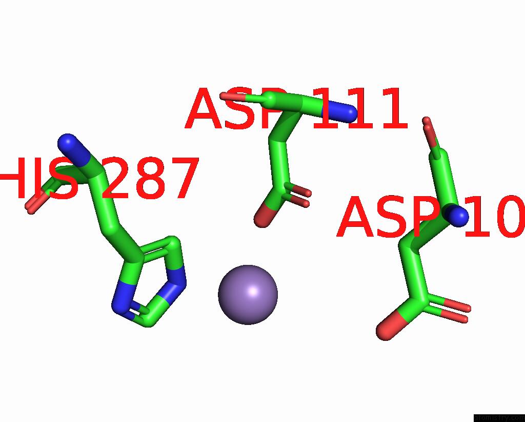
Manganese binding site 1 out of 2 in 8hl8
Go back to
Manganese binding site 1 out
of 2 in the Crystal Structrue of Mtdl R257K Mutant
Mono view
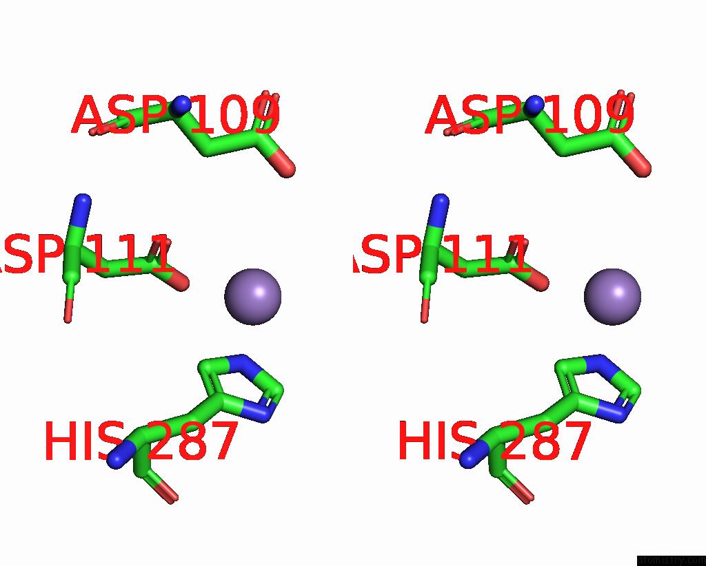
Stereo pair view
Mono view
Stereo pair view
A full contact list of Manganese with other atoms in the Mn binding
site number 1 of Crystal Structrue of Mtdl R257K Mutant within 5.0Å range:
|
Manganese binding site 2 out of 2 in 8hl8
Go back to
Manganese binding site 2 out
of 2 in the Crystal Structrue of Mtdl R257K Mutant

Mono view
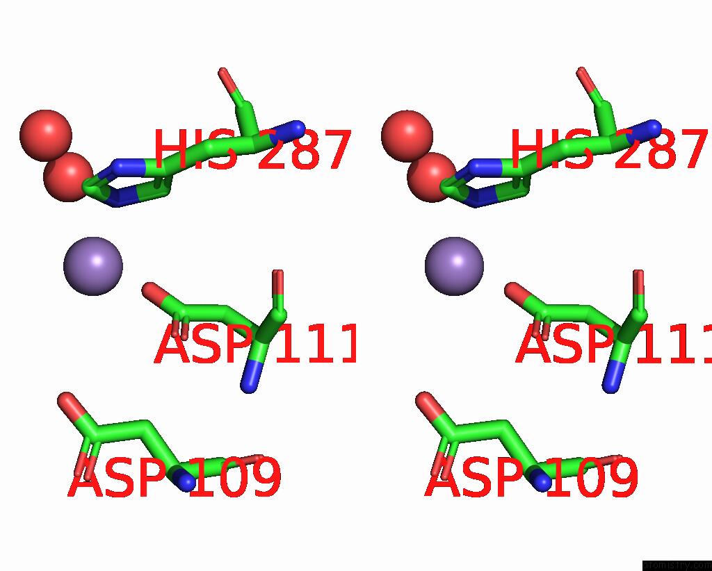
Stereo pair view
Mono view
Stereo pair view
A full contact list of Manganese with other atoms in the Mn binding
site number 2 of Crystal Structrue of Mtdl R257K Mutant within 5.0Å range:
|
Reference:
X.Du,
X.Chu,
N.Liu,
X.Jia,
H.Peng,
Y.Xiao,
L.Liu,
H.Yu,
F.Li,
C.He.
Structures of the Ndp-Pyranose Mutase Belonging to Glycosyltransferase Family 75 Reveal Residues Important For Mn 2+ Coordination and Substrate Binding. J.Biol.Chem. V. 299 02903 2023.
ISSN: ESSN 1083-351X
PubMed: 36642179
DOI: 10.1016/J.JBC.2023.102903
Page generated: Sun Oct 6 12:28:45 2024
ISSN: ESSN 1083-351X
PubMed: 36642179
DOI: 10.1016/J.JBC.2023.102903
Last articles
Zn in 9MJ5Zn in 9HNW
Zn in 9G0L
Zn in 9FNE
Zn in 9DZN
Zn in 9E0I
Zn in 9D32
Zn in 9DAK
Zn in 8ZXC
Zn in 8ZUF