Manganese »
PDB 8f4d-8hl8 »
8hbb »
Manganese in PDB 8hbb: Crystal Structure of Caenorhabditis Elegans Nmad-1 in Complex with Ligand III
Enzymatic activity of Crystal Structure of Caenorhabditis Elegans Nmad-1 in Complex with Ligand III
All present enzymatic activity of Crystal Structure of Caenorhabditis Elegans Nmad-1 in Complex with Ligand III:
1.14.11.51;
1.14.11.51;
Protein crystallography data
The structure of Crystal Structure of Caenorhabditis Elegans Nmad-1 in Complex with Ligand III, PDB code: 8hbb
was solved by
G.Shang,
Z.Chen,
with X-Ray Crystallography technique. A brief refinement statistics is given in the table below:
| Resolution Low / High (Å) | 47.18 / 3.09 |
| Space group | C 1 2 1 |
| Cell size a, b, c (Å), α, β, γ (°) | 183.176, 75.328, 118.184, 90, 113.19, 90 |
| R / Rfree (%) | 26 / 28.2 |
Other elements in 8hbb:
The structure of Crystal Structure of Caenorhabditis Elegans Nmad-1 in Complex with Ligand III also contains other interesting chemical elements:
| Chlorine | (Cl) | 4 atoms |
Manganese Binding Sites:
The binding sites of Manganese atom in the Crystal Structure of Caenorhabditis Elegans Nmad-1 in Complex with Ligand III
(pdb code 8hbb). This binding sites where shown within
5.0 Angstroms radius around Manganese atom.
In total 5 binding sites of Manganese where determined in the Crystal Structure of Caenorhabditis Elegans Nmad-1 in Complex with Ligand III, PDB code: 8hbb:
Jump to Manganese binding site number: 1; 2; 3; 4; 5;
In total 5 binding sites of Manganese where determined in the Crystal Structure of Caenorhabditis Elegans Nmad-1 in Complex with Ligand III, PDB code: 8hbb:
Jump to Manganese binding site number: 1; 2; 3; 4; 5;
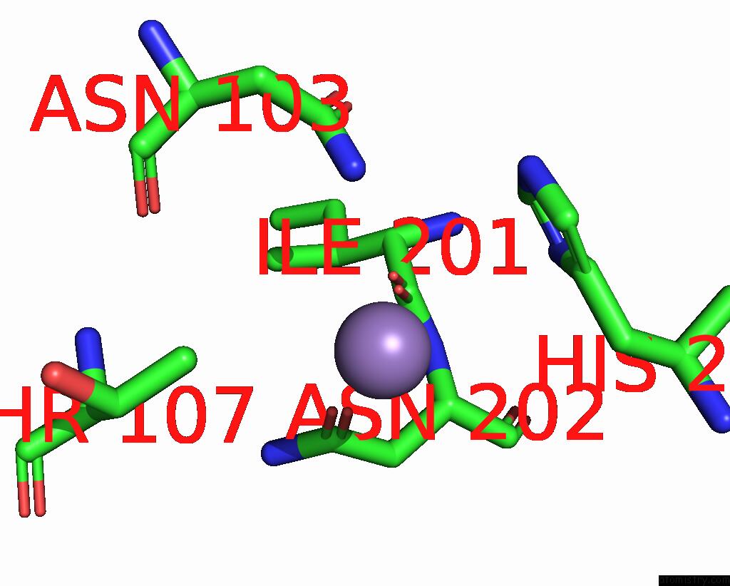
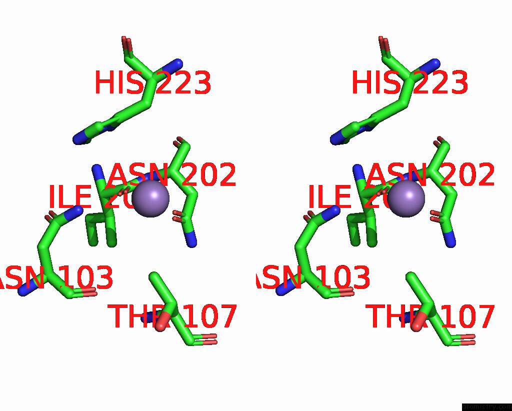
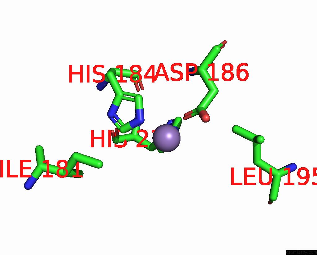
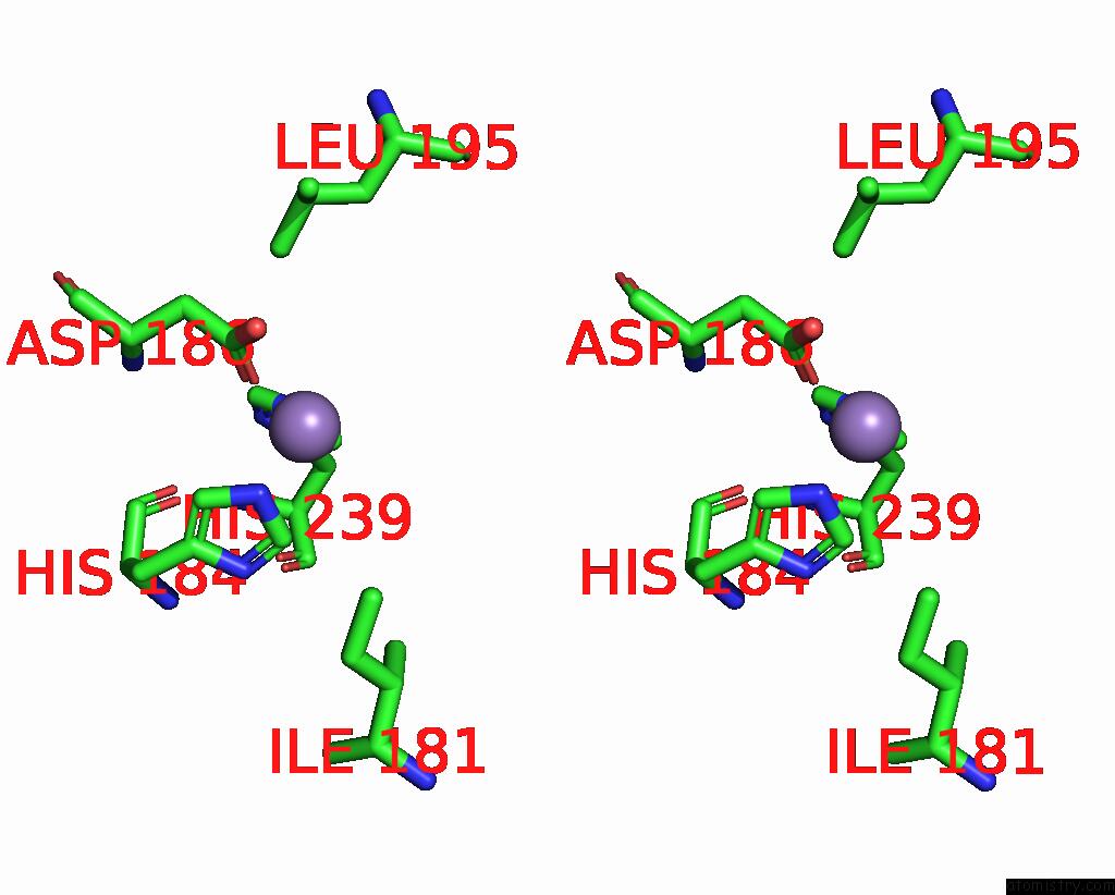
Manganese binding site 1 out of 5 in 8hbb
Go back to
Manganese binding site 1 out
of 5 in the Crystal Structure of Caenorhabditis Elegans Nmad-1 in Complex with Ligand III
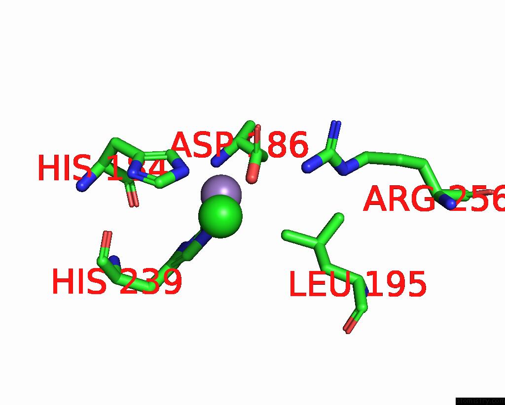
Mono view
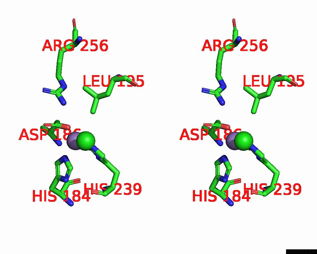
Stereo pair view
Mono view
Stereo pair view
A full contact list of Manganese with other atoms in the Mn binding
site number 1 of Crystal Structure of Caenorhabditis Elegans Nmad-1 in Complex with Ligand III within 5.0Å range:
|
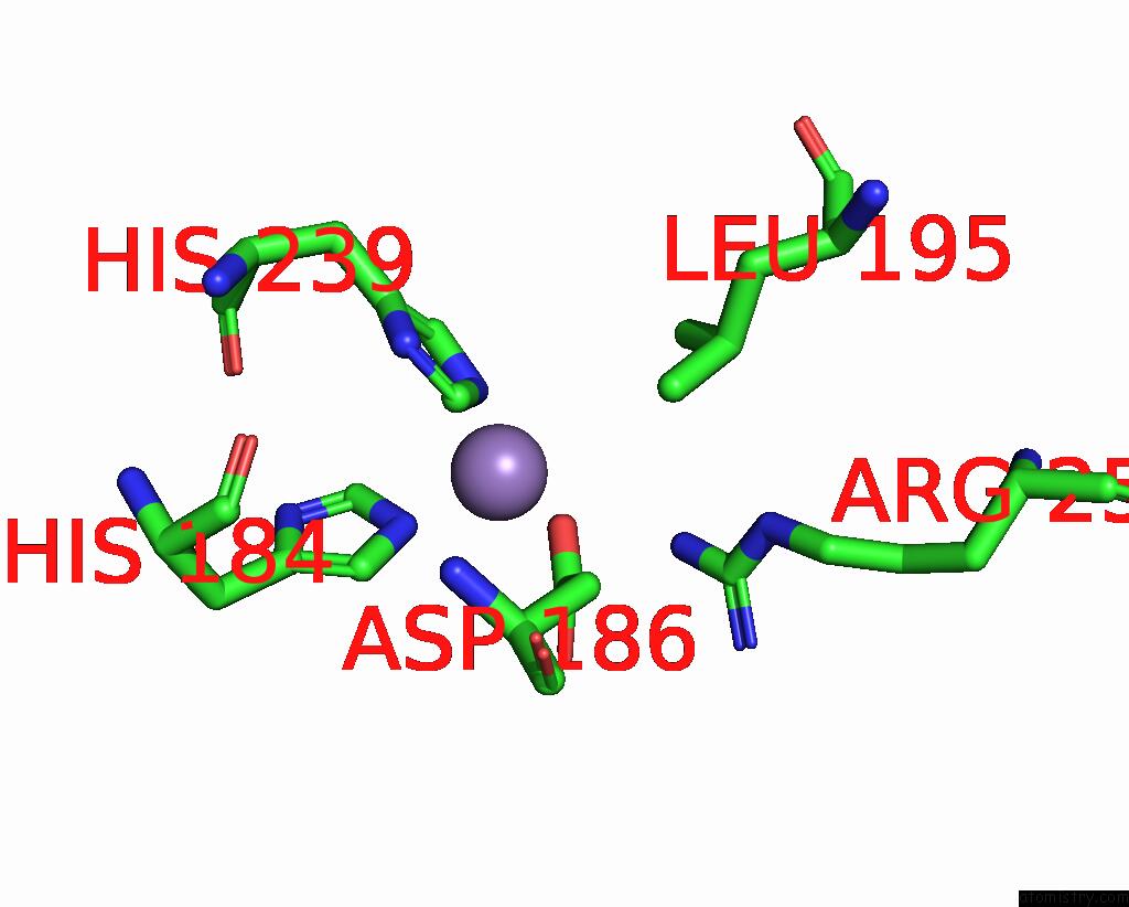
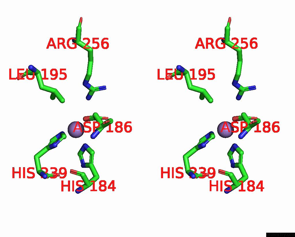
Manganese binding site 2 out of 5 in 8hbb
Go back to
Manganese binding site 2 out
of 5 in the Crystal Structure of Caenorhabditis Elegans Nmad-1 in Complex with Ligand III
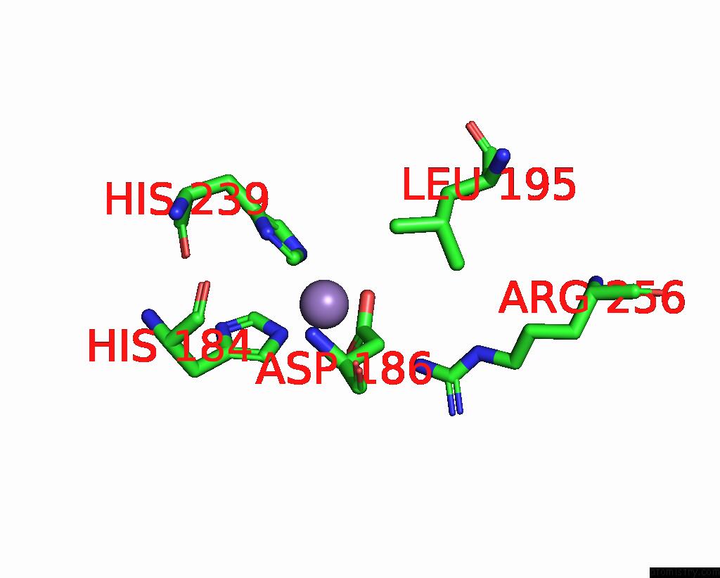
Mono view
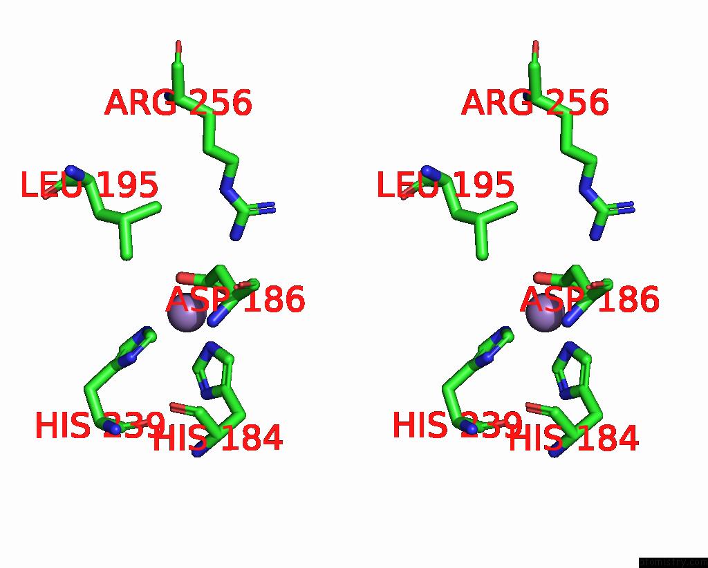
Stereo pair view
Mono view
Stereo pair view
A full contact list of Manganese with other atoms in the Mn binding
site number 2 of Crystal Structure of Caenorhabditis Elegans Nmad-1 in Complex with Ligand III within 5.0Å range:
|
Manganese binding site 3 out of 5 in 8hbb
Go back to
Manganese binding site 3 out
of 5 in the Crystal Structure of Caenorhabditis Elegans Nmad-1 in Complex with Ligand III
Mono view
Stereo pair view
Mono view
Stereo pair view
A full contact list of Manganese with other atoms in the Mn binding
site number 3 of Crystal Structure of Caenorhabditis Elegans Nmad-1 in Complex with Ligand III within 5.0Å range:
|
Manganese binding site 4 out of 5 in 8hbb
Go back to
Manganese binding site 4 out
of 5 in the Crystal Structure of Caenorhabditis Elegans Nmad-1 in Complex with Ligand III
Mono view
Stereo pair view
Mono view
Stereo pair view
A full contact list of Manganese with other atoms in the Mn binding
site number 4 of Crystal Structure of Caenorhabditis Elegans Nmad-1 in Complex with Ligand III within 5.0Å range:
|
Manganese binding site 5 out of 5 in 8hbb
Go back to
Manganese binding site 5 out
of 5 in the Crystal Structure of Caenorhabditis Elegans Nmad-1 in Complex with Ligand III
Mono view
Stereo pair view
Mono view
Stereo pair view
A full contact list of Manganese with other atoms in the Mn binding
site number 5 of Crystal Structure of Caenorhabditis Elegans Nmad-1 in Complex with Ligand III within 5.0Å range:
|
Reference:
G.Shang,
M.Yang,
M.Li,
L.Ma,
Y.Liu,
J.Ma,
Y.Chen,
X.Wang,
S.Fan,
M.Xie,
W.Wu,
S.Dai,
Z.Chen.
Structural Basis of Nucleic Acid Recognition and 6MA Demethylation By Caenorhabditis Elegans Nmad-1A. Int J Mol Sci V. 25 2024.
ISSN: ESSN 1422-0067
PubMed: 38255759
DOI: 10.3390/IJMS25020686
Page generated: Sun Oct 6 12:27:13 2024
ISSN: ESSN 1422-0067
PubMed: 38255759
DOI: 10.3390/IJMS25020686
Last articles
Zn in 9MJ5Zn in 9HNW
Zn in 9G0L
Zn in 9FNE
Zn in 9DZN
Zn in 9E0I
Zn in 9D32
Zn in 9DAK
Zn in 8ZXC
Zn in 8ZUF