Manganese »
PDB 8f4d-8hl8 »
8h2c »
Manganese in PDB 8h2c: Crystal Structure of the Pseudaminic Acid Synthase Psei From Campylobacter Jejuni
Enzymatic activity of Crystal Structure of the Pseudaminic Acid Synthase Psei From Campylobacter Jejuni
All present enzymatic activity of Crystal Structure of the Pseudaminic Acid Synthase Psei From Campylobacter Jejuni:
2.5.1.97;
2.5.1.97;
Protein crystallography data
The structure of Crystal Structure of the Pseudaminic Acid Synthase Psei From Campylobacter Jejuni, PDB code: 8h2c
was solved by
W.S.Song,
M.A.Park,
D.U.Ki,
S.I.Yoon,
with X-Ray Crystallography technique. A brief refinement statistics is given in the table below:
| Resolution Low / High (Å) | 30.00 / 2.90 |
| Space group | P 4 21 2 |
| Cell size a, b, c (Å), α, β, γ (°) | 176.805, 176.805, 99.824, 90, 90, 90 |
| R / Rfree (%) | 24.5 / 29.5 |
Manganese Binding Sites:
The binding sites of Manganese atom in the Crystal Structure of the Pseudaminic Acid Synthase Psei From Campylobacter Jejuni
(pdb code 8h2c). This binding sites where shown within
5.0 Angstroms radius around Manganese atom.
In total 4 binding sites of Manganese where determined in the Crystal Structure of the Pseudaminic Acid Synthase Psei From Campylobacter Jejuni, PDB code: 8h2c:
Jump to Manganese binding site number: 1; 2; 3; 4;
In total 4 binding sites of Manganese where determined in the Crystal Structure of the Pseudaminic Acid Synthase Psei From Campylobacter Jejuni, PDB code: 8h2c:
Jump to Manganese binding site number: 1; 2; 3; 4;
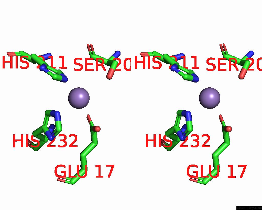
Manganese binding site 1 out of 4 in 8h2c
Go back to
Manganese binding site 1 out
of 4 in the Crystal Structure of the Pseudaminic Acid Synthase Psei From Campylobacter Jejuni
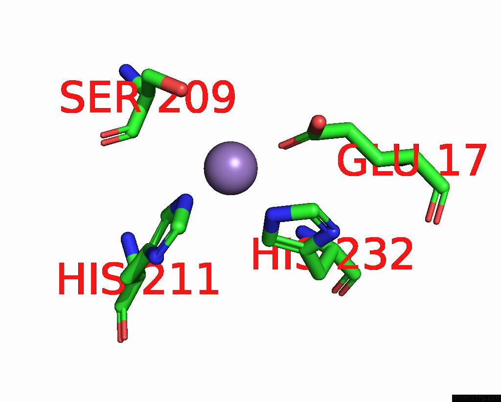
Mono view
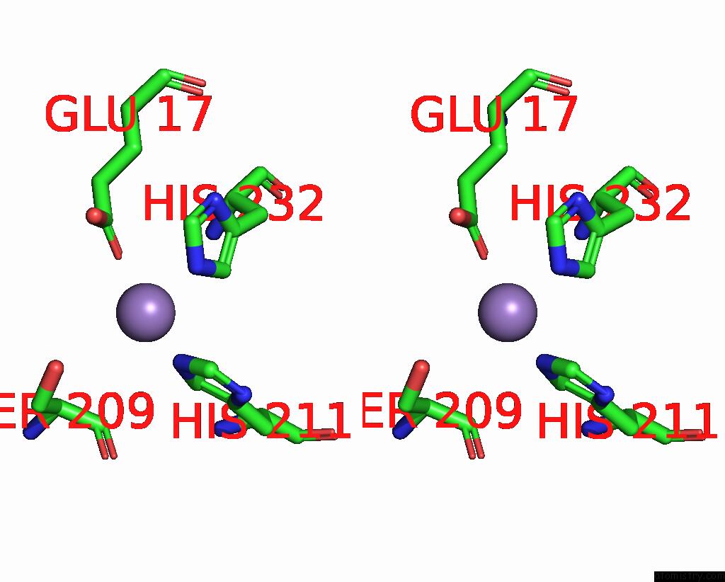
Stereo pair view
Mono view
Stereo pair view
A full contact list of Manganese with other atoms in the Mn binding
site number 1 of Crystal Structure of the Pseudaminic Acid Synthase Psei From Campylobacter Jejuni within 5.0Å range:
|
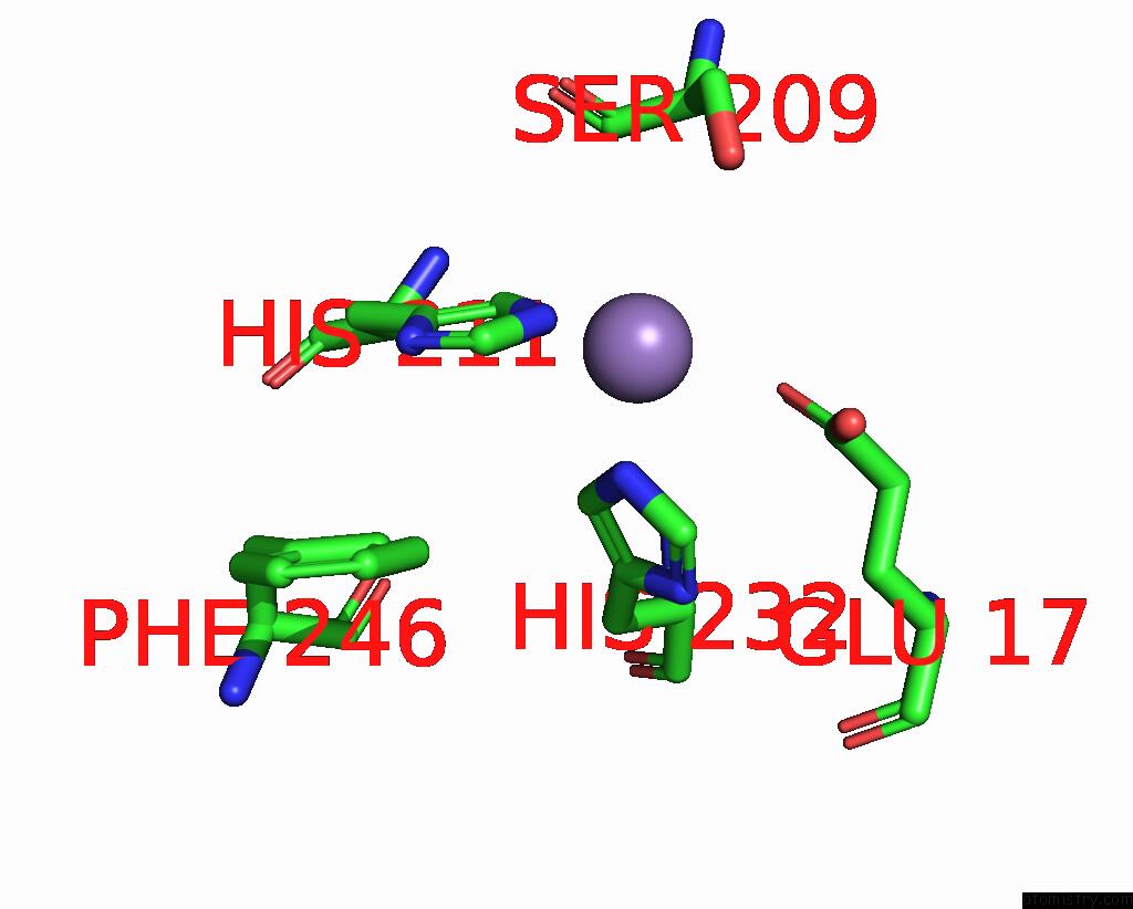
Manganese binding site 2 out of 4 in 8h2c
Go back to
Manganese binding site 2 out
of 4 in the Crystal Structure of the Pseudaminic Acid Synthase Psei From Campylobacter Jejuni
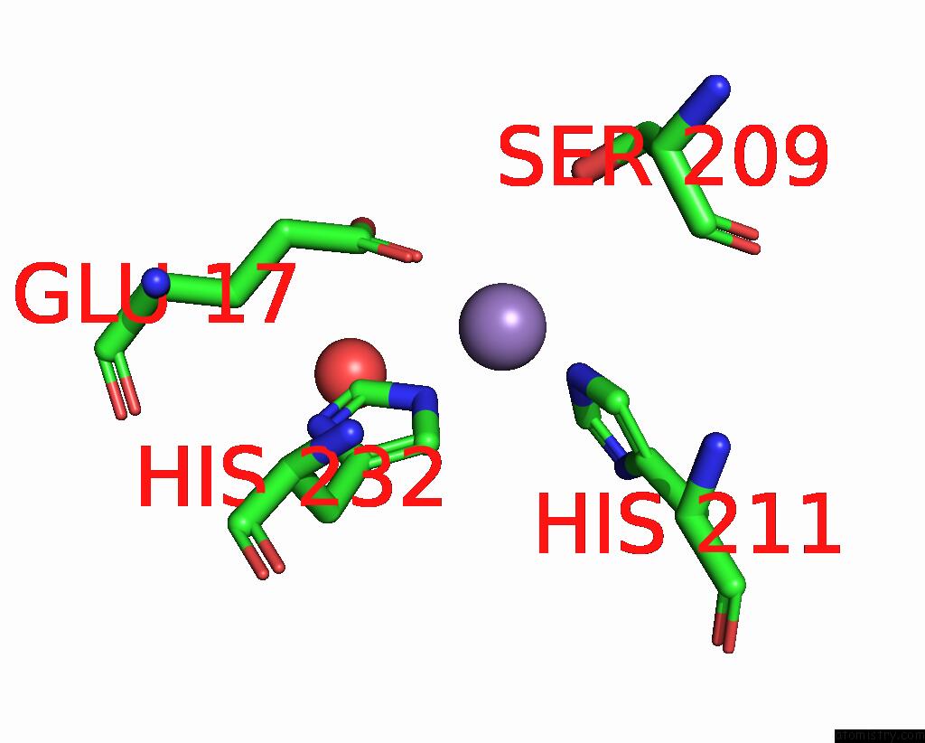
Mono view
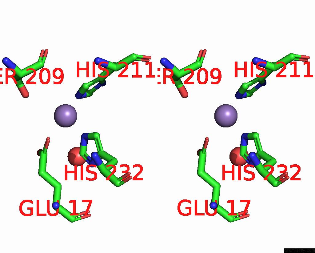
Stereo pair view
Mono view
Stereo pair view
A full contact list of Manganese with other atoms in the Mn binding
site number 2 of Crystal Structure of the Pseudaminic Acid Synthase Psei From Campylobacter Jejuni within 5.0Å range:
|
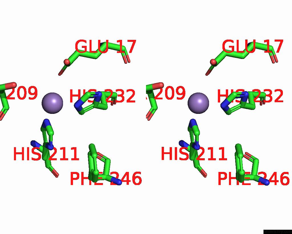
Manganese binding site 3 out of 4 in 8h2c
Go back to
Manganese binding site 3 out
of 4 in the Crystal Structure of the Pseudaminic Acid Synthase Psei From Campylobacter Jejuni

Mono view
Stereo pair view
Mono view
Stereo pair view
A full contact list of Manganese with other atoms in the Mn binding
site number 3 of Crystal Structure of the Pseudaminic Acid Synthase Psei From Campylobacter Jejuni within 5.0Å range:
|
Manganese binding site 4 out of 4 in 8h2c
Go back to
Manganese binding site 4 out
of 4 in the Crystal Structure of the Pseudaminic Acid Synthase Psei From Campylobacter Jejuni
Mono view
Stereo pair view
Mono view
Stereo pair view
A full contact list of Manganese with other atoms in the Mn binding
site number 4 of Crystal Structure of the Pseudaminic Acid Synthase Psei From Campylobacter Jejuni within 5.0Å range:
|
Reference:
W.S.Song,
M.A.Park,
D.U.Ki,
S.I.Yoon.
Structural Analysis of the Pseudaminic Acid Synthase Psei From Campylobacter Jejuni. Biochem.Biophys.Res.Commun. V. 635 252 2022.
ISSN: ESSN 1090-2104
PubMed: 36283338
DOI: 10.1016/J.BBRC.2022.10.050
Page generated: Sun Aug 17 00:58:06 2025
ISSN: ESSN 1090-2104
PubMed: 36283338
DOI: 10.1016/J.BBRC.2022.10.050
Last articles
Na in 2AP1Na in 2AMF
Na in 2AHR
Na in 2AHY
Na in 2AOC
Na in 2ANP
Na in 2AGV
Na in 2AHS
Na in 2AER
Na in 2AFH