Manganese »
PDB 8f4d-8hl8 »
8ffd »
Manganese in PDB 8ffd: Crystal Structure of Manganeese Bound Dps Protein (PA0962) From Pseudomonas Aeruginosa (Cubic Form)
Protein crystallography data
The structure of Crystal Structure of Manganeese Bound Dps Protein (PA0962) From Pseudomonas Aeruginosa (Cubic Form), PDB code: 8ffd
was solved by
S.Lovell,
S.Seibold,
K.P.Battaile,
M.Rivera,
with X-Ray Crystallography technique. A brief refinement statistics is given in the table below:
| Resolution Low / High (Å) | 48.86 / 2.20 |
| Space group | P 21 3 1 |
| Cell size a, b, c (Å), α, β, γ (°) | 223.914, 223.914, 223.914, 90, 90, 90 |
| R / Rfree (%) | 17 / 22.4 |
Manganese Binding Sites:
Pages:
>>> Page 1 <<< Page 2, Binding sites: 11 - 16;Binding sites:
The binding sites of Manganese atom in the Crystal Structure of Manganeese Bound Dps Protein (PA0962) From Pseudomonas Aeruginosa (Cubic Form) (pdb code 8ffd). This binding sites where shown within 5.0 Angstroms radius around Manganese atom.In total 16 binding sites of Manganese where determined in the Crystal Structure of Manganeese Bound Dps Protein (PA0962) From Pseudomonas Aeruginosa (Cubic Form), PDB code: 8ffd:
Jump to Manganese binding site number: 1; 2; 3; 4; 5; 6; 7; 8; 9; 10;
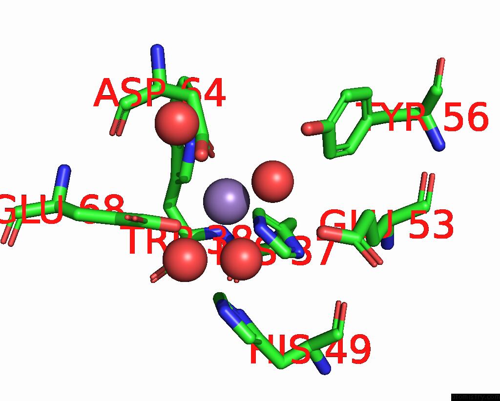
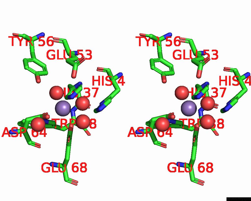
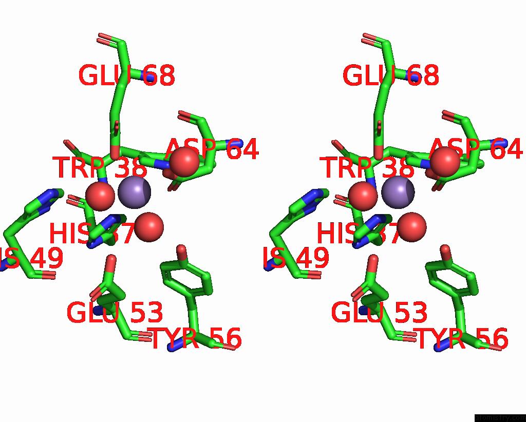
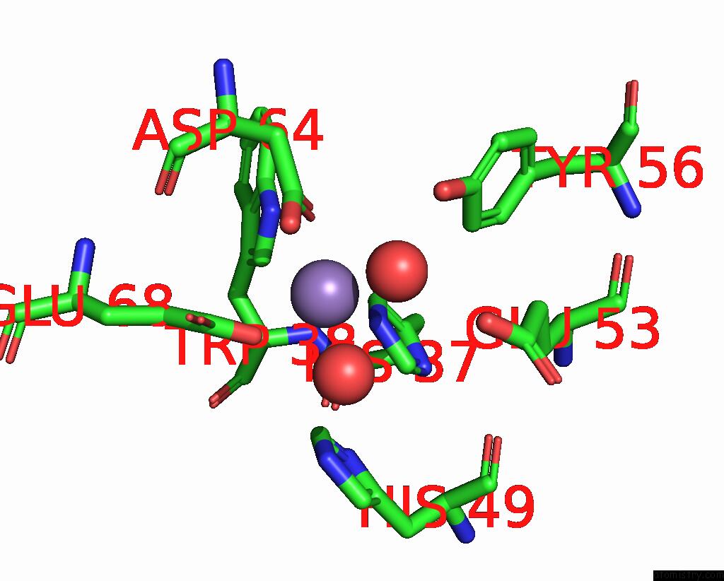
Manganese binding site 1 out of 16 in 8ffd
Go back to
Manganese binding site 1 out
of 16 in the Crystal Structure of Manganeese Bound Dps Protein (PA0962) From Pseudomonas Aeruginosa (Cubic Form)
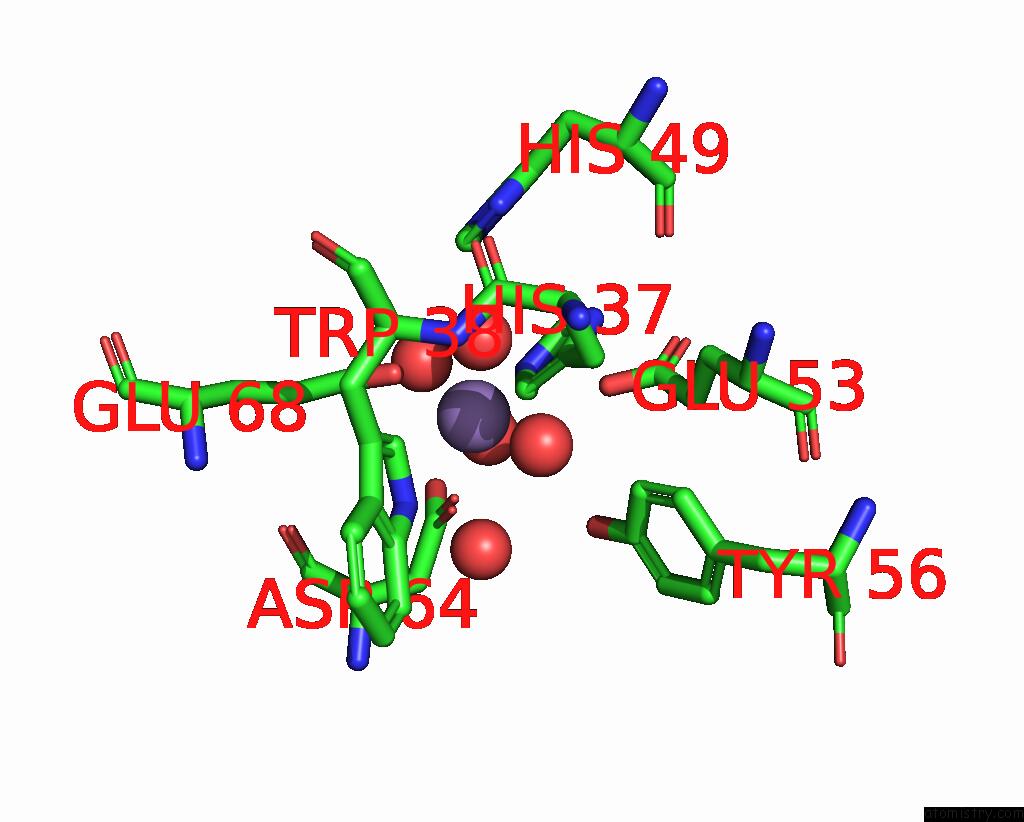
Mono view
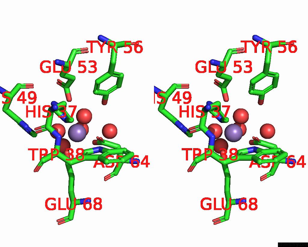
Stereo pair view
Mono view
Stereo pair view
A full contact list of Manganese with other atoms in the Mn binding
site number 1 of Crystal Structure of Manganeese Bound Dps Protein (PA0962) From Pseudomonas Aeruginosa (Cubic Form) within 5.0Å range:
|
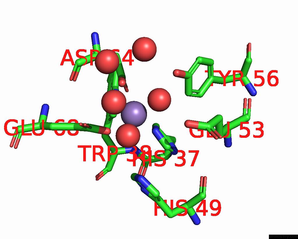
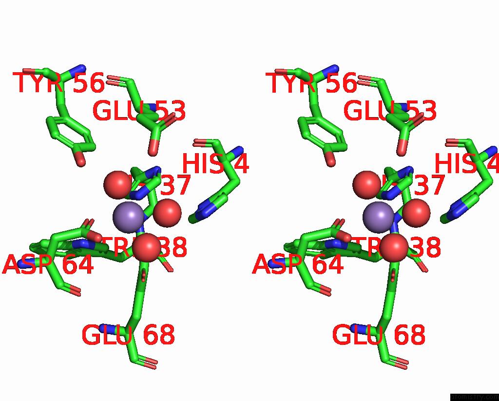
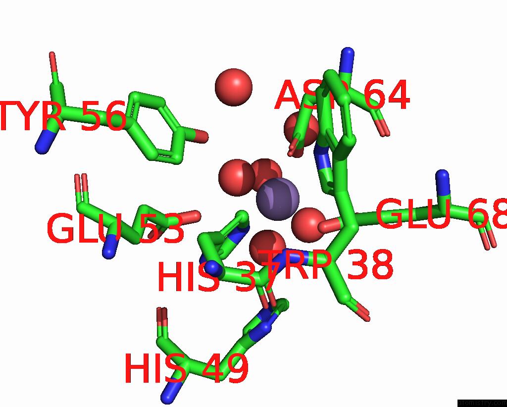
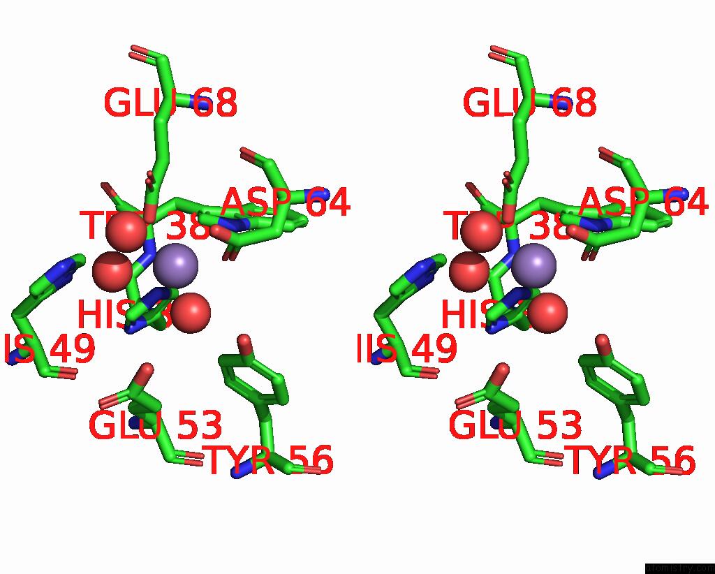
Manganese binding site 2 out of 16 in 8ffd
Go back to
Manganese binding site 2 out
of 16 in the Crystal Structure of Manganeese Bound Dps Protein (PA0962) From Pseudomonas Aeruginosa (Cubic Form)
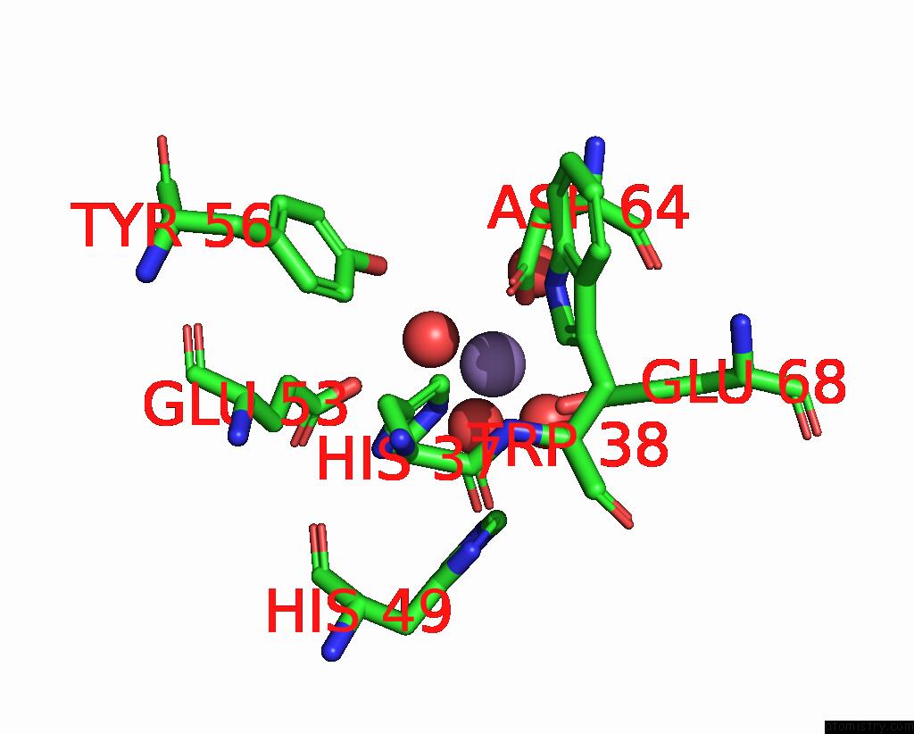
Mono view
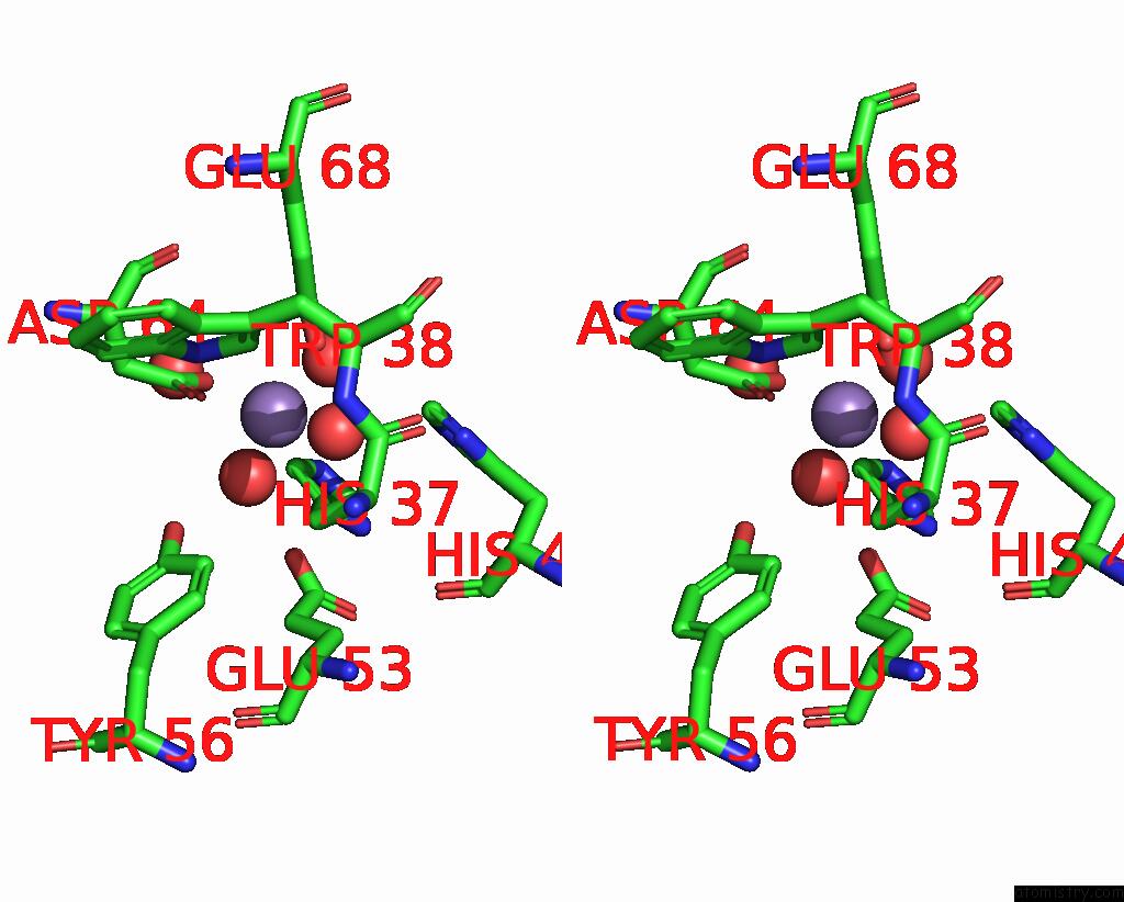
Stereo pair view
Mono view
Stereo pair view
A full contact list of Manganese with other atoms in the Mn binding
site number 2 of Crystal Structure of Manganeese Bound Dps Protein (PA0962) From Pseudomonas Aeruginosa (Cubic Form) within 5.0Å range:
|
Manganese binding site 3 out of 16 in 8ffd
Go back to
Manganese binding site 3 out
of 16 in the Crystal Structure of Manganeese Bound Dps Protein (PA0962) From Pseudomonas Aeruginosa (Cubic Form)
Mono view
Stereo pair view
Mono view
Stereo pair view
A full contact list of Manganese with other atoms in the Mn binding
site number 3 of Crystal Structure of Manganeese Bound Dps Protein (PA0962) From Pseudomonas Aeruginosa (Cubic Form) within 5.0Å range:
|
Manganese binding site 4 out of 16 in 8ffd
Go back to
Manganese binding site 4 out
of 16 in the Crystal Structure of Manganeese Bound Dps Protein (PA0962) From Pseudomonas Aeruginosa (Cubic Form)
Mono view

Stereo pair view
Mono view
Stereo pair view
A full contact list of Manganese with other atoms in the Mn binding
site number 4 of Crystal Structure of Manganeese Bound Dps Protein (PA0962) From Pseudomonas Aeruginosa (Cubic Form) within 5.0Å range:
|
Manganese binding site 5 out of 16 in 8ffd
Go back to
Manganese binding site 5 out
of 16 in the Crystal Structure of Manganeese Bound Dps Protein (PA0962) From Pseudomonas Aeruginosa (Cubic Form)

Mono view
Stereo pair view
Mono view
Stereo pair view
A full contact list of Manganese with other atoms in the Mn binding
site number 5 of Crystal Structure of Manganeese Bound Dps Protein (PA0962) From Pseudomonas Aeruginosa (Cubic Form) within 5.0Å range:
|
Manganese binding site 6 out of 16 in 8ffd
Go back to
Manganese binding site 6 out
of 16 in the Crystal Structure of Manganeese Bound Dps Protein (PA0962) From Pseudomonas Aeruginosa (Cubic Form)

Mono view
Stereo pair view
Mono view
Stereo pair view
A full contact list of Manganese with other atoms in the Mn binding
site number 6 of Crystal Structure of Manganeese Bound Dps Protein (PA0962) From Pseudomonas Aeruginosa (Cubic Form) within 5.0Å range:
|
Manganese binding site 7 out of 16 in 8ffd
Go back to
Manganese binding site 7 out
of 16 in the Crystal Structure of Manganeese Bound Dps Protein (PA0962) From Pseudomonas Aeruginosa (Cubic Form)
Mono view

Stereo pair view
Mono view
Stereo pair view
A full contact list of Manganese with other atoms in the Mn binding
site number 7 of Crystal Structure of Manganeese Bound Dps Protein (PA0962) From Pseudomonas Aeruginosa (Cubic Form) within 5.0Å range:
|
Manganese binding site 8 out of 16 in 8ffd
Go back to
Manganese binding site 8 out
of 16 in the Crystal Structure of Manganeese Bound Dps Protein (PA0962) From Pseudomonas Aeruginosa (Cubic Form)
Mono view

Stereo pair view
Mono view
Stereo pair view
A full contact list of Manganese with other atoms in the Mn binding
site number 8 of Crystal Structure of Manganeese Bound Dps Protein (PA0962) From Pseudomonas Aeruginosa (Cubic Form) within 5.0Å range:
|
Manganese binding site 9 out of 16 in 8ffd
Go back to
Manganese binding site 9 out
of 16 in the Crystal Structure of Manganeese Bound Dps Protein (PA0962) From Pseudomonas Aeruginosa (Cubic Form)

Mono view

Stereo pair view
Mono view
Stereo pair view
A full contact list of Manganese with other atoms in the Mn binding
site number 9 of Crystal Structure of Manganeese Bound Dps Protein (PA0962) From Pseudomonas Aeruginosa (Cubic Form) within 5.0Å range:
|
Manganese binding site 10 out of 16 in 8ffd
Go back to
Manganese binding site 10 out
of 16 in the Crystal Structure of Manganeese Bound Dps Protein (PA0962) From Pseudomonas Aeruginosa (Cubic Form)

Mono view
Stereo pair view
Mono view
Stereo pair view
A full contact list of Manganese with other atoms in the Mn binding
site number 10 of Crystal Structure of Manganeese Bound Dps Protein (PA0962) From Pseudomonas Aeruginosa (Cubic Form) within 5.0Å range:
|
Reference:
N.Rajapaksha,
A.Soldano,
H.Yao,
F.Donnarumma,
M.M.Kashipathy,
S.Seibold,
K.P.Battaile,
S.Lovell,
M.Rivera.
Pseudomonas Aeruginosa Dps (PA0962) Functions in H2O2 Mediated Oxidative Stress Defense and Exhibits in Vitro Dna Cleaving Activity Int J Mol Sci V. 24 2023.
ISSN: ESSN 1422-0067
DOI: 10.3390/IJMS24054669
Page generated: Sun Oct 6 12:06:57 2024
ISSN: ESSN 1422-0067
DOI: 10.3390/IJMS24054669
Last articles
Cl in 7TMUCl in 7TJP
Cl in 7TMZ
Cl in 7TLG
Cl in 7TJC
Cl in 7TJO
Cl in 7TLE
Cl in 7TKV
Cl in 7THH
Cl in 7TIV