Manganese »
PDB 8dl1-8f4c »
8eip »
Manganese in PDB 8eip: Crystal Structure of Cyanophycin Dipeptide Hydrolase Cphz E251A From Acinetobacter Baylyi DSM587 in Complex with Beta-Asp-Arg
Protein crystallography data
The structure of Crystal Structure of Cyanophycin Dipeptide Hydrolase Cphz E251A From Acinetobacter Baylyi DSM587 in Complex with Beta-Asp-Arg, PDB code: 8eip
was solved by
I.Sharon,
T.M.Schmeing,
with X-Ray Crystallography technique. A brief refinement statistics is given in the table below:
| Resolution Low / High (Å) | 49.04 / 2.24 |
| Space group | C 1 2 1 |
| Cell size a, b, c (Å), α, β, γ (°) | 151.529, 126.648, 109.516, 90, 130.39, 90 |
| R / Rfree (%) | 22.6 / 25.8 |
Other elements in 8eip:
The structure of Crystal Structure of Cyanophycin Dipeptide Hydrolase Cphz E251A From Acinetobacter Baylyi DSM587 in Complex with Beta-Asp-Arg also contains other interesting chemical elements:
| Zinc | (Zn) | 4 atoms |
Manganese Binding Sites:
The binding sites of Manganese atom in the Crystal Structure of Cyanophycin Dipeptide Hydrolase Cphz E251A From Acinetobacter Baylyi DSM587 in Complex with Beta-Asp-Arg
(pdb code 8eip). This binding sites where shown within
5.0 Angstroms radius around Manganese atom.
In total 4 binding sites of Manganese where determined in the Crystal Structure of Cyanophycin Dipeptide Hydrolase Cphz E251A From Acinetobacter Baylyi DSM587 in Complex with Beta-Asp-Arg, PDB code: 8eip:
Jump to Manganese binding site number: 1; 2; 3; 4;
In total 4 binding sites of Manganese where determined in the Crystal Structure of Cyanophycin Dipeptide Hydrolase Cphz E251A From Acinetobacter Baylyi DSM587 in Complex with Beta-Asp-Arg, PDB code: 8eip:
Jump to Manganese binding site number: 1; 2; 3; 4;
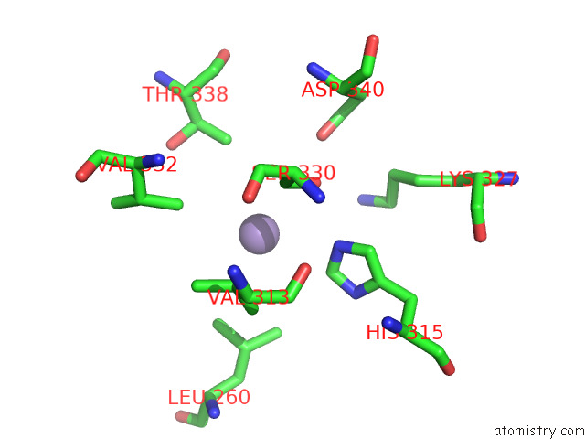
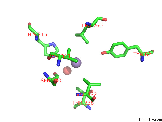
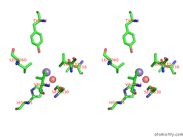
Manganese binding site 1 out of 4 in 8eip
Go back to
Manganese binding site 1 out
of 4 in the Crystal Structure of Cyanophycin Dipeptide Hydrolase Cphz E251A From Acinetobacter Baylyi DSM587 in Complex with Beta-Asp-Arg
Mono view
Stereo pair view
Mono view
Stereo pair view
A full contact list of Manganese with other atoms in the Mn binding
site number 1 of Crystal Structure of Cyanophycin Dipeptide Hydrolase Cphz E251A From Acinetobacter Baylyi DSM587 in Complex with Beta-Asp-Arg within 5.0Å range:
|
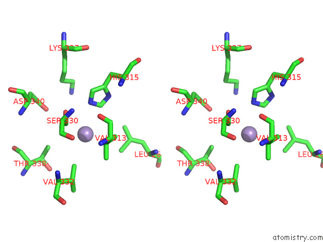
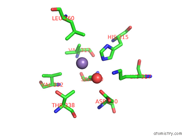
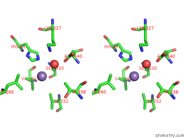
Manganese binding site 2 out of 4 in 8eip
Go back to
Manganese binding site 2 out
of 4 in the Crystal Structure of Cyanophycin Dipeptide Hydrolase Cphz E251A From Acinetobacter Baylyi DSM587 in Complex with Beta-Asp-Arg
Mono view
Stereo pair view
Mono view
Stereo pair view
A full contact list of Manganese with other atoms in the Mn binding
site number 2 of Crystal Structure of Cyanophycin Dipeptide Hydrolase Cphz E251A From Acinetobacter Baylyi DSM587 in Complex with Beta-Asp-Arg within 5.0Å range:
|
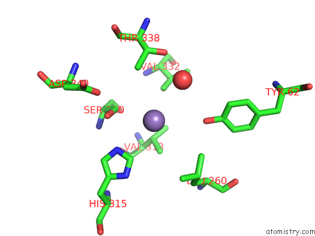
Manganese binding site 3 out of 4 in 8eip
Go back to
Manganese binding site 3 out
of 4 in the Crystal Structure of Cyanophycin Dipeptide Hydrolase Cphz E251A From Acinetobacter Baylyi DSM587 in Complex with Beta-Asp-Arg
Mono view
Stereo pair view
Mono view
Stereo pair view
A full contact list of Manganese with other atoms in the Mn binding
site number 3 of Crystal Structure of Cyanophycin Dipeptide Hydrolase Cphz E251A From Acinetobacter Baylyi DSM587 in Complex with Beta-Asp-Arg within 5.0Å range:
|
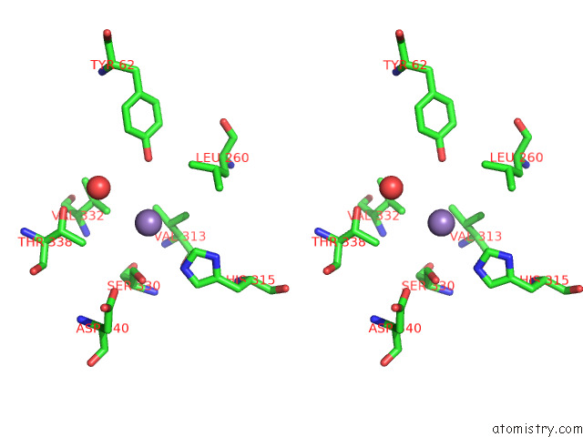
Manganese binding site 4 out of 4 in 8eip
Go back to
Manganese binding site 4 out
of 4 in the Crystal Structure of Cyanophycin Dipeptide Hydrolase Cphz E251A From Acinetobacter Baylyi DSM587 in Complex with Beta-Asp-Arg
Mono view
Stereo pair view
Mono view
Stereo pair view
A full contact list of Manganese with other atoms in the Mn binding
site number 4 of Crystal Structure of Cyanophycin Dipeptide Hydrolase Cphz E251A From Acinetobacter Baylyi DSM587 in Complex with Beta-Asp-Arg within 5.0Å range:
|
Reference:
I.Sharon,
G.A.Mckay,
D.Nguyen,
T.M.Schmeing.
Discovery of Cyanophycin Dipeptide Hydrolase Enzymes Suggests Widespread Utility of the Natural Biopolymer Cyanophycin. Proc.Natl.Acad.Sci.Usa V. 120 47120 2023.
ISSN: ESSN 1091-6490
PubMed: 36800389
DOI: 10.1073/PNAS.2216547120
Page generated: Sun Oct 6 11:42:17 2024
ISSN: ESSN 1091-6490
PubMed: 36800389
DOI: 10.1073/PNAS.2216547120
Last articles
Zn in 9MJ5Zn in 9HNW
Zn in 9G0L
Zn in 9FNE
Zn in 9DZN
Zn in 9E0I
Zn in 9D32
Zn in 9DAK
Zn in 8ZXC
Zn in 8ZUF