Manganese »
PDB 8dl1-8f4c »
8dq4 »
Manganese in PDB 8dq4: X-Ray Crystal Structure of Flavobacterium Johnsoniae Dimanganese(II) Class Id Ribonucleotide Reductase Beta Subunit K71R Variant
Enzymatic activity of X-Ray Crystal Structure of Flavobacterium Johnsoniae Dimanganese(II) Class Id Ribonucleotide Reductase Beta Subunit K71R Variant
All present enzymatic activity of X-Ray Crystal Structure of Flavobacterium Johnsoniae Dimanganese(II) Class Id Ribonucleotide Reductase Beta Subunit K71R Variant:
1.17.4.1;
1.17.4.1;
Protein crystallography data
The structure of X-Ray Crystal Structure of Flavobacterium Johnsoniae Dimanganese(II) Class Id Ribonucleotide Reductase Beta Subunit K71R Variant, PDB code: 8dq4
was solved by
H.R.Rose,
A.O.Maggiolo,
J.J.Jung,
A.K.Boal,
with X-Ray Crystallography technique. A brief refinement statistics is given in the table below:
| Resolution Low / High (Å) | 28.84 / 2.35 |
| Space group | P 31 |
| Cell size a, b, c (Å), α, β, γ (°) | 53.551, 53.551, 220.947, 90, 90, 120 |
| R / Rfree (%) | 22.7 / 27.5 |
Manganese Binding Sites:
The binding sites of Manganese atom in the X-Ray Crystal Structure of Flavobacterium Johnsoniae Dimanganese(II) Class Id Ribonucleotide Reductase Beta Subunit K71R Variant
(pdb code 8dq4). This binding sites where shown within
5.0 Angstroms radius around Manganese atom.
In total 4 binding sites of Manganese where determined in the X-Ray Crystal Structure of Flavobacterium Johnsoniae Dimanganese(II) Class Id Ribonucleotide Reductase Beta Subunit K71R Variant, PDB code: 8dq4:
Jump to Manganese binding site number: 1; 2; 3; 4;
In total 4 binding sites of Manganese where determined in the X-Ray Crystal Structure of Flavobacterium Johnsoniae Dimanganese(II) Class Id Ribonucleotide Reductase Beta Subunit K71R Variant, PDB code: 8dq4:
Jump to Manganese binding site number: 1; 2; 3; 4;
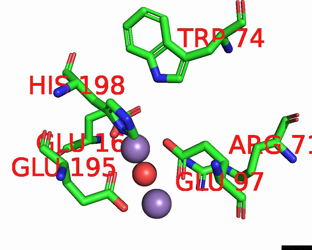
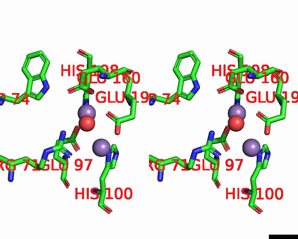
Manganese binding site 1 out of 4 in 8dq4
Go back to
Manganese binding site 1 out
of 4 in the X-Ray Crystal Structure of Flavobacterium Johnsoniae Dimanganese(II) Class Id Ribonucleotide Reductase Beta Subunit K71R Variant
Mono view
Stereo pair view
Mono view
Stereo pair view
A full contact list of Manganese with other atoms in the Mn binding
site number 1 of X-Ray Crystal Structure of Flavobacterium Johnsoniae Dimanganese(II) Class Id Ribonucleotide Reductase Beta Subunit K71R Variant within 5.0Å range:
|
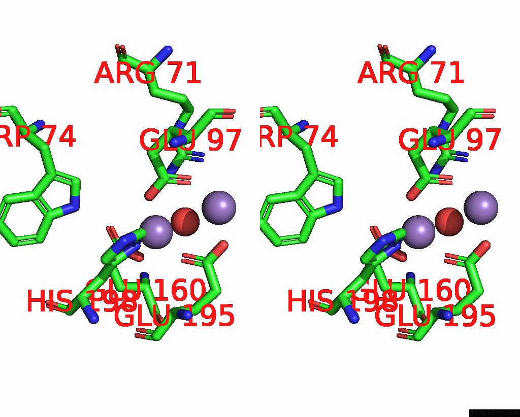
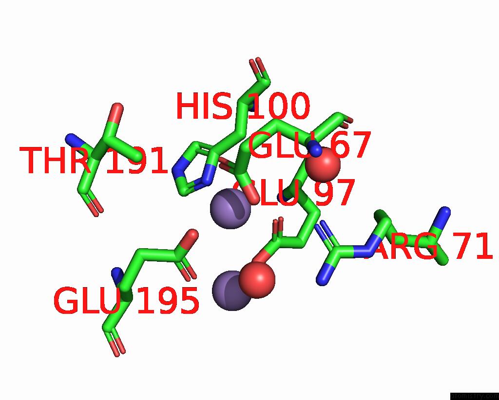
Manganese binding site 2 out of 4 in 8dq4
Go back to
Manganese binding site 2 out
of 4 in the X-Ray Crystal Structure of Flavobacterium Johnsoniae Dimanganese(II) Class Id Ribonucleotide Reductase Beta Subunit K71R Variant

Mono view
Stereo pair view
Mono view
Stereo pair view
A full contact list of Manganese with other atoms in the Mn binding
site number 2 of X-Ray Crystal Structure of Flavobacterium Johnsoniae Dimanganese(II) Class Id Ribonucleotide Reductase Beta Subunit K71R Variant within 5.0Å range:
|
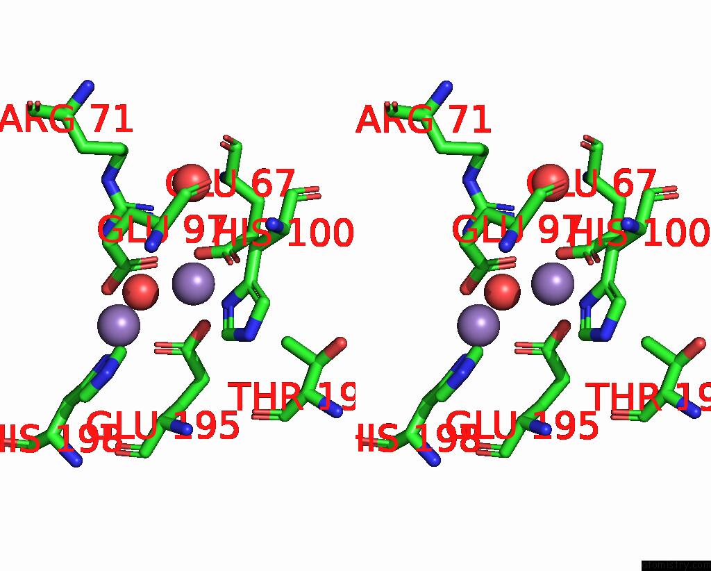
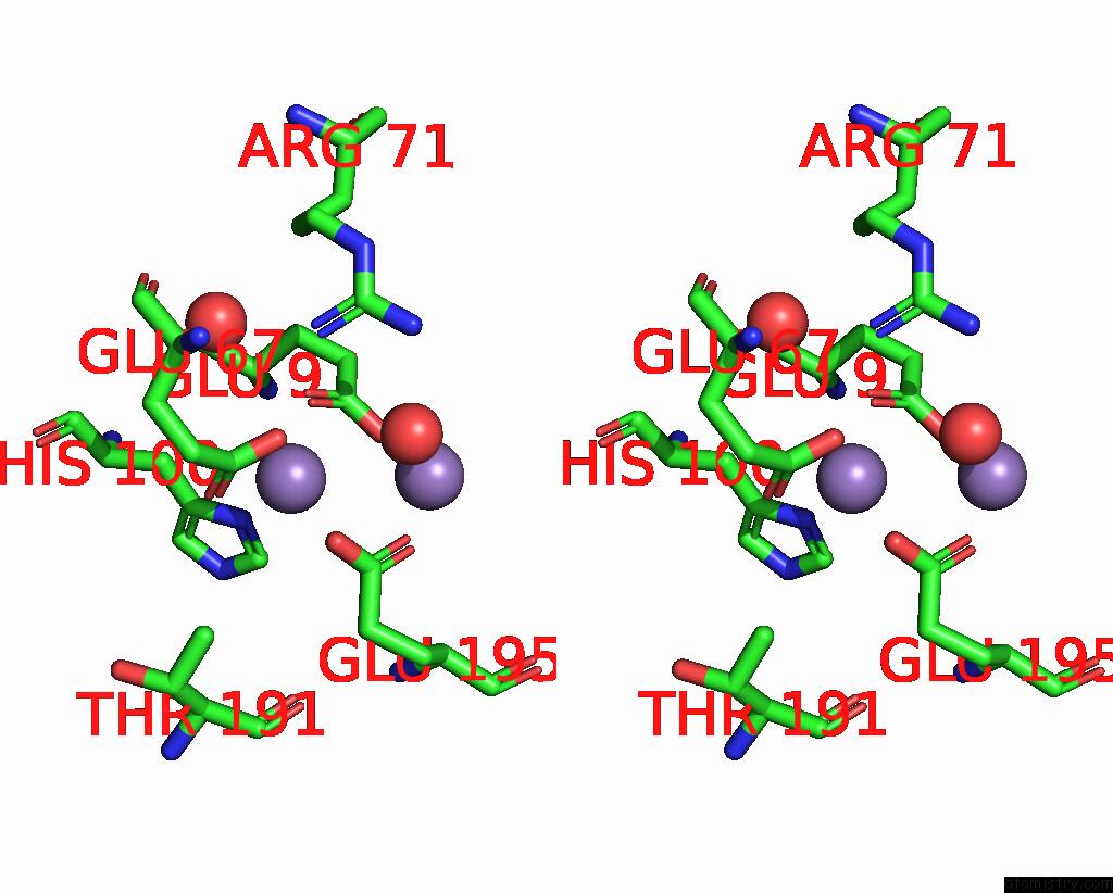
Manganese binding site 3 out of 4 in 8dq4
Go back to
Manganese binding site 3 out
of 4 in the X-Ray Crystal Structure of Flavobacterium Johnsoniae Dimanganese(II) Class Id Ribonucleotide Reductase Beta Subunit K71R Variant
Mono view
Stereo pair view
Mono view
Stereo pair view
A full contact list of Manganese with other atoms in the Mn binding
site number 3 of X-Ray Crystal Structure of Flavobacterium Johnsoniae Dimanganese(II) Class Id Ribonucleotide Reductase Beta Subunit K71R Variant within 5.0Å range:
|
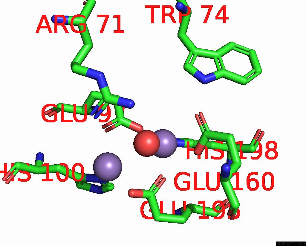
Manganese binding site 4 out of 4 in 8dq4
Go back to
Manganese binding site 4 out
of 4 in the X-Ray Crystal Structure of Flavobacterium Johnsoniae Dimanganese(II) Class Id Ribonucleotide Reductase Beta Subunit K71R Variant
Mono view
Stereo pair view
Mono view
Stereo pair view
A full contact list of Manganese with other atoms in the Mn binding
site number 4 of X-Ray Crystal Structure of Flavobacterium Johnsoniae Dimanganese(II) Class Id Ribonucleotide Reductase Beta Subunit K71R Variant within 5.0Å range:
|
Reference:
H.R.Rose,
A.K.Boal.
X-Ray Crystal Structure of Flavobacterium Johnsoniae Dimanganese(II) Class Id Ribonucleotide Reductase Beta Subunit K71R Variant To Be Published.
Page generated: Sun Oct 6 11:33:45 2024
Last articles
Cl in 7ZYDCl in 7ZXV
Cl in 7ZWY
Cl in 7ZX4
Cl in 7ZXD
Cl in 7ZX2
Cl in 7ZWX
Cl in 7ZWT
Cl in 7ZWU
Cl in 7ZW2