Manganese »
PDB 8awx-8djy »
8bvk »
Manganese in PDB 8bvk: The Crystal Structure of O-Glycoside Cleaving Beta-Eliminase From A. Tumefaciens Atoge
Protein crystallography data
The structure of The Crystal Structure of O-Glycoside Cleaving Beta-Eliminase From A. Tumefaciens Atoge, PDB code: 8bvk
was solved by
K.Kuhlmann,
J.Bitter,
M.Pfeiffer,
B.Nidetzky,
T.Pavkov-Keller,
with X-Ray Crystallography technique. A brief refinement statistics is given in the table below:
| Resolution Low / High (Å) | 46.84 / 2.00 |
| Space group | P 21 21 21 |
| Cell size a, b, c (Å), α, β, γ (°) | 36.58, 93.67, 123.68, 90, 90, 90 |
| R / Rfree (%) | 17.4 / 21.8 |
Manganese Binding Sites:
The binding sites of Manganese atom in the The Crystal Structure of O-Glycoside Cleaving Beta-Eliminase From A. Tumefaciens Atoge
(pdb code 8bvk). This binding sites where shown within
5.0 Angstroms radius around Manganese atom.
In total 3 binding sites of Manganese where determined in the The Crystal Structure of O-Glycoside Cleaving Beta-Eliminase From A. Tumefaciens Atoge, PDB code: 8bvk:
Jump to Manganese binding site number: 1; 2; 3;
In total 3 binding sites of Manganese where determined in the The Crystal Structure of O-Glycoside Cleaving Beta-Eliminase From A. Tumefaciens Atoge, PDB code: 8bvk:
Jump to Manganese binding site number: 1; 2; 3;
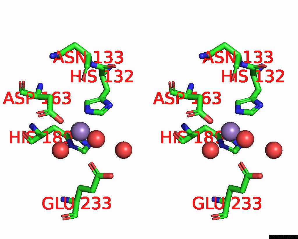
Manganese binding site 1 out of 3 in 8bvk
Go back to
Manganese binding site 1 out
of 3 in the The Crystal Structure of O-Glycoside Cleaving Beta-Eliminase From A. Tumefaciens Atoge
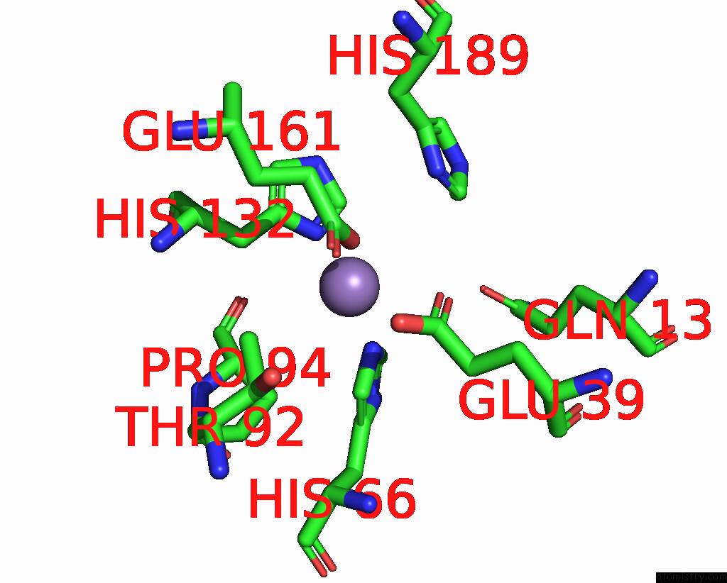
Mono view
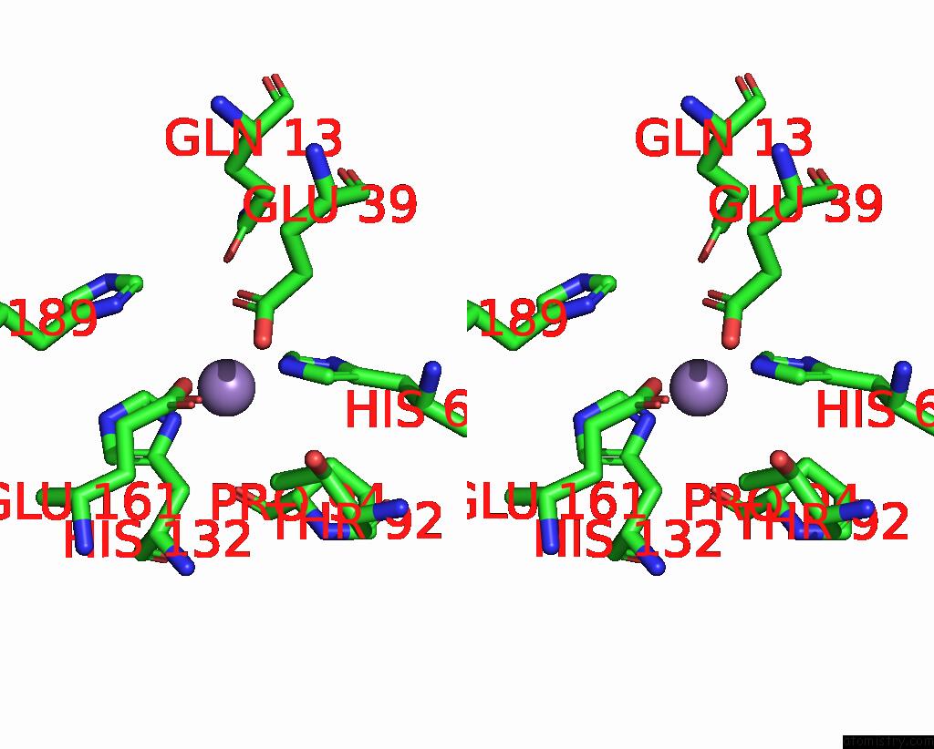
Stereo pair view
Mono view
Stereo pair view
A full contact list of Manganese with other atoms in the Mn binding
site number 1 of The Crystal Structure of O-Glycoside Cleaving Beta-Eliminase From A. Tumefaciens Atoge within 5.0Å range:
|
Manganese binding site 2 out of 3 in 8bvk
Go back to
Manganese binding site 2 out
of 3 in the The Crystal Structure of O-Glycoside Cleaving Beta-Eliminase From A. Tumefaciens Atoge
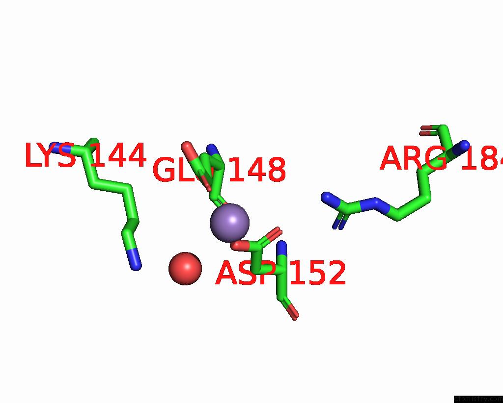
Mono view

Stereo pair view
Mono view
Stereo pair view
A full contact list of Manganese with other atoms in the Mn binding
site number 2 of The Crystal Structure of O-Glycoside Cleaving Beta-Eliminase From A. Tumefaciens Atoge within 5.0Å range:
|
Manganese binding site 3 out of 3 in 8bvk
Go back to
Manganese binding site 3 out
of 3 in the The Crystal Structure of O-Glycoside Cleaving Beta-Eliminase From A. Tumefaciens Atoge
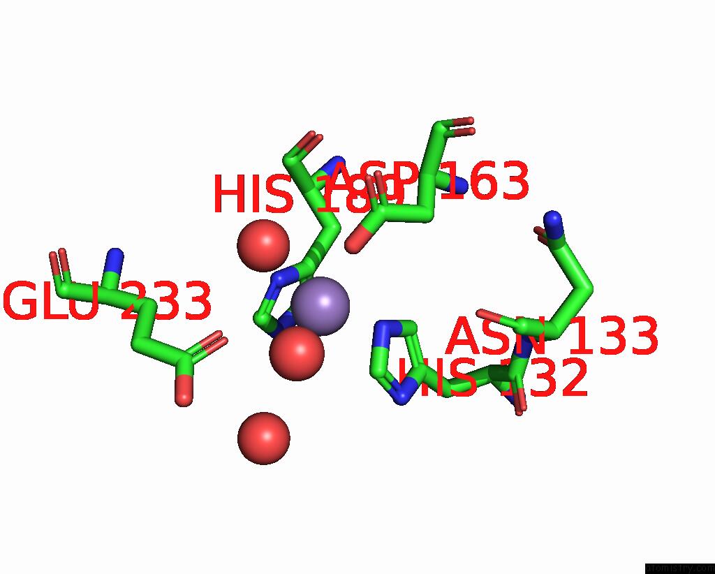
Mono view
Stereo pair view
Mono view
Stereo pair view
A full contact list of Manganese with other atoms in the Mn binding
site number 3 of The Crystal Structure of O-Glycoside Cleaving Beta-Eliminase From A. Tumefaciens Atoge within 5.0Å range:
|
Reference:
J.Bitter,
M.Pfeiffer,
A.J.E.Borg,
K.Kuhlmann,
T.Pavkov-Keller,
B.Nidetzky.
Enzymatic Beta-Elimination in Natural Product Deglycosylation: C-C Bond Cleavage As A Specialty of A Conserved Mechanism To Be Published.
Page generated: Sun Oct 6 11:27:31 2024
Last articles
Zn in 9J0NZn in 9J0O
Zn in 9J0P
Zn in 9FJX
Zn in 9EKB
Zn in 9C0F
Zn in 9CAH
Zn in 9CH0
Zn in 9CH3
Zn in 9CH1