Manganese »
PDB 7z03-8awv »
7z3e »
Manganese in PDB 7z3e: Xfel Structure of Class Ib Ribonucleotide Reductase Dimanganese(II) Nrdf in Complex with Hydroquinone Nrdi From Bacillus Cereus
Enzymatic activity of Xfel Structure of Class Ib Ribonucleotide Reductase Dimanganese(II) Nrdf in Complex with Hydroquinone Nrdi From Bacillus Cereus
All present enzymatic activity of Xfel Structure of Class Ib Ribonucleotide Reductase Dimanganese(II) Nrdf in Complex with Hydroquinone Nrdi From Bacillus Cereus:
1.17.4.1;
1.17.4.1;
Protein crystallography data
The structure of Xfel Structure of Class Ib Ribonucleotide Reductase Dimanganese(II) Nrdf in Complex with Hydroquinone Nrdi From Bacillus Cereus, PDB code: 7z3e
was solved by
J.John,
H.Lebrette,
O.Aurelius,
M.Hogbom,
with X-Ray Crystallography technique. A brief refinement statistics is given in the table below:
| Resolution Low / High (Å) | 23.91 / 2.00 |
| Space group | C 2 2 21 |
| Cell size a, b, c (Å), α, β, γ (°) | 61.237, 125.795, 144.77, 90, 90, 90 |
| R / Rfree (%) | 15.2 / 18.4 |
Manganese Binding Sites:
The binding sites of Manganese atom in the Xfel Structure of Class Ib Ribonucleotide Reductase Dimanganese(II) Nrdf in Complex with Hydroquinone Nrdi From Bacillus Cereus
(pdb code 7z3e). This binding sites where shown within
5.0 Angstroms radius around Manganese atom.
In total 2 binding sites of Manganese where determined in the Xfel Structure of Class Ib Ribonucleotide Reductase Dimanganese(II) Nrdf in Complex with Hydroquinone Nrdi From Bacillus Cereus, PDB code: 7z3e:
Jump to Manganese binding site number: 1; 2;
In total 2 binding sites of Manganese where determined in the Xfel Structure of Class Ib Ribonucleotide Reductase Dimanganese(II) Nrdf in Complex with Hydroquinone Nrdi From Bacillus Cereus, PDB code: 7z3e:
Jump to Manganese binding site number: 1; 2;
Manganese binding site 1 out of 2 in 7z3e
Go back to
Manganese binding site 1 out
of 2 in the Xfel Structure of Class Ib Ribonucleotide Reductase Dimanganese(II) Nrdf in Complex with Hydroquinone Nrdi From Bacillus Cereus
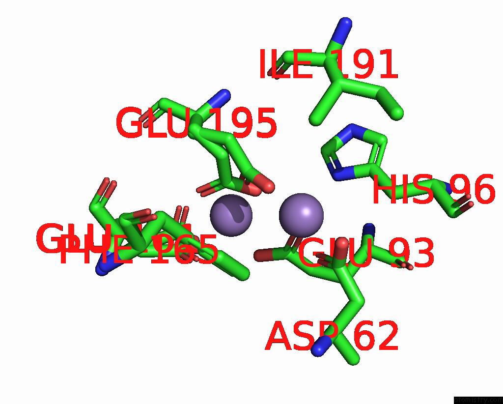
Mono view
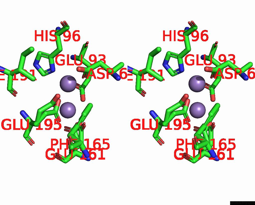
Stereo pair view
Mono view
Stereo pair view
A full contact list of Manganese with other atoms in the Mn binding
site number 1 of Xfel Structure of Class Ib Ribonucleotide Reductase Dimanganese(II) Nrdf in Complex with Hydroquinone Nrdi From Bacillus Cereus within 5.0Å range:
|
Manganese binding site 2 out of 2 in 7z3e
Go back to
Manganese binding site 2 out
of 2 in the Xfel Structure of Class Ib Ribonucleotide Reductase Dimanganese(II) Nrdf in Complex with Hydroquinone Nrdi From Bacillus Cereus
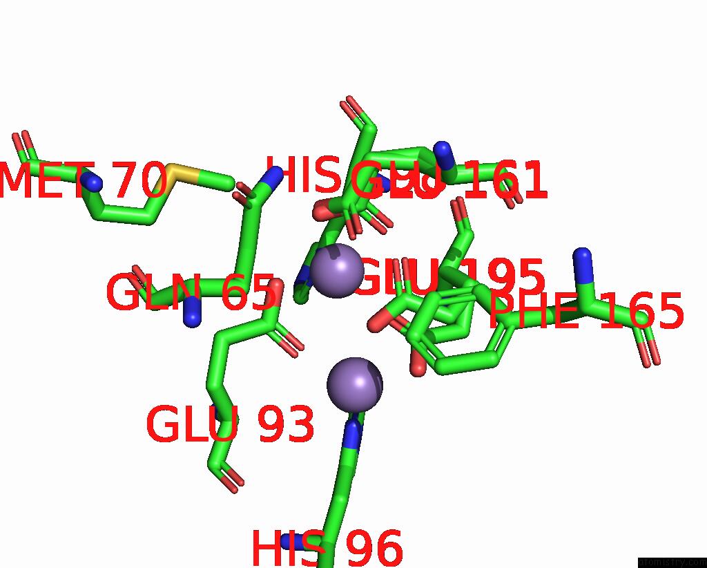
Mono view
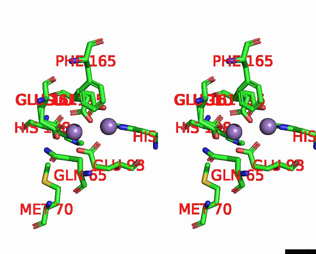
Stereo pair view
Mono view
Stereo pair view
A full contact list of Manganese with other atoms in the Mn binding
site number 2 of Xfel Structure of Class Ib Ribonucleotide Reductase Dimanganese(II) Nrdf in Complex with Hydroquinone Nrdi From Bacillus Cereus within 5.0Å range:
|
Reference:
J.John,
O.Aurelius,
V.Srinivas,
P.Saura,
I.S.Kim,
A.Bhowmick,
P.S.Simon,
M.Dasgupta,
C.Pham,
S.Gul,
K.D.Sutherlin,
P.Aller,
A.Butryn,
A.M.Orville,
M.H.Cheah,
S.Owada,
K.Tono,
F.D.Fuller,
A.Batyuk,
A.S.Brewster,
N.K.Sauter,
V.K.Yachandra,
J.Yano,
V.R.I.Kaila,
J.Kern,
H.Lebrette,
M.Hogbom.
Redox-Controlled Reorganization and Flavin Strain Within the Ribonucleotide Reductase R2B-Nrdi Complex Monitored By Serial Femtosecond Crystallography. Elife V. 11 2022.
ISSN: ESSN 2050-084X
PubMed: 36083619
DOI: 10.7554/ELIFE.79226
Page generated: Sun Oct 6 11:13:49 2024
ISSN: ESSN 2050-084X
PubMed: 36083619
DOI: 10.7554/ELIFE.79226
Last articles
Zn in 9J0NZn in 9J0O
Zn in 9J0P
Zn in 9FJX
Zn in 9EKB
Zn in 9C0F
Zn in 9CAH
Zn in 9CH0
Zn in 9CH3
Zn in 9CH1