Manganese »
PDB 7x9j-7yzp »
7xiu »
Manganese in PDB 7xiu: Crystal Structure of Engineered Hiv-1 Reverse Transcriptase Rnase H Domain Complexed with Nitrofuran Methoxy(Methoxycarbonyl)Phenyl Ester
Enzymatic activity of Crystal Structure of Engineered Hiv-1 Reverse Transcriptase Rnase H Domain Complexed with Nitrofuran Methoxy(Methoxycarbonyl)Phenyl Ester
All present enzymatic activity of Crystal Structure of Engineered Hiv-1 Reverse Transcriptase Rnase H Domain Complexed with Nitrofuran Methoxy(Methoxycarbonyl)Phenyl Ester:
3.1.26.13;
3.1.26.13;
Protein crystallography data
The structure of Crystal Structure of Engineered Hiv-1 Reverse Transcriptase Rnase H Domain Complexed with Nitrofuran Methoxy(Methoxycarbonyl)Phenyl Ester, PDB code: 7xiu
was solved by
H.Lu,
Y.Komukai,
K.Usami,
Y.Guo,
X.Qiao,
M.Nukaga,
T.Hoshino,
with X-Ray Crystallography technique. A brief refinement statistics is given in the table below:
| Resolution Low / High (Å) | 43.88 / 2.09 |
| Space group | P 41 21 2 |
| Cell size a, b, c (Å), α, β, γ (°) | 62.061, 62.061, 83.001, 90, 90, 90 |
| R / Rfree (%) | 22.1 / 26.7 |
Other elements in 7xiu:
The structure of Crystal Structure of Engineered Hiv-1 Reverse Transcriptase Rnase H Domain Complexed with Nitrofuran Methoxy(Methoxycarbonyl)Phenyl Ester also contains other interesting chemical elements:
| Zinc | (Zn) | 2 atoms |
Manganese Binding Sites:
The binding sites of Manganese atom in the Crystal Structure of Engineered Hiv-1 Reverse Transcriptase Rnase H Domain Complexed with Nitrofuran Methoxy(Methoxycarbonyl)Phenyl Ester
(pdb code 7xiu). This binding sites where shown within
5.0 Angstroms radius around Manganese atom.
In total 2 binding sites of Manganese where determined in the Crystal Structure of Engineered Hiv-1 Reverse Transcriptase Rnase H Domain Complexed with Nitrofuran Methoxy(Methoxycarbonyl)Phenyl Ester, PDB code: 7xiu:
Jump to Manganese binding site number: 1; 2;
In total 2 binding sites of Manganese where determined in the Crystal Structure of Engineered Hiv-1 Reverse Transcriptase Rnase H Domain Complexed with Nitrofuran Methoxy(Methoxycarbonyl)Phenyl Ester, PDB code: 7xiu:
Jump to Manganese binding site number: 1; 2;
Manganese binding site 1 out of 2 in 7xiu
Go back to
Manganese binding site 1 out
of 2 in the Crystal Structure of Engineered Hiv-1 Reverse Transcriptase Rnase H Domain Complexed with Nitrofuran Methoxy(Methoxycarbonyl)Phenyl Ester
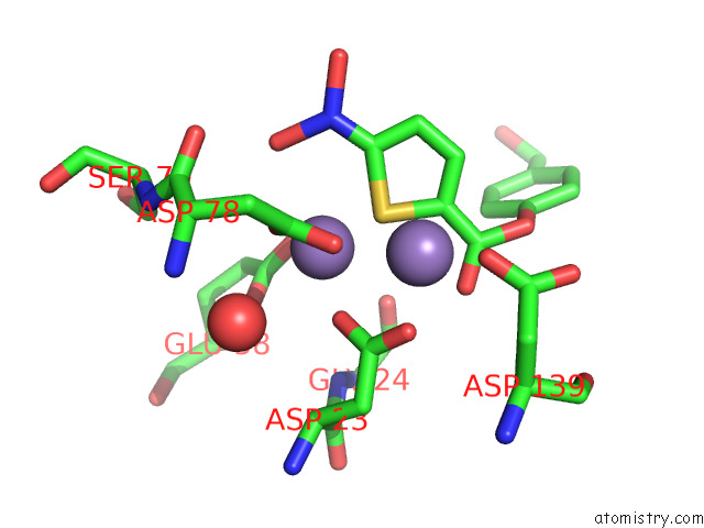
Mono view
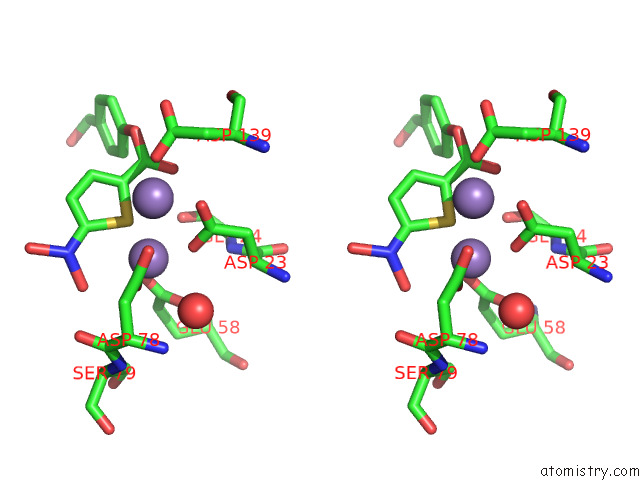
Stereo pair view
Mono view
Stereo pair view
A full contact list of Manganese with other atoms in the Mn binding
site number 1 of Crystal Structure of Engineered Hiv-1 Reverse Transcriptase Rnase H Domain Complexed with Nitrofuran Methoxy(Methoxycarbonyl)Phenyl Ester within 5.0Å range:
|
Manganese binding site 2 out of 2 in 7xiu
Go back to
Manganese binding site 2 out
of 2 in the Crystal Structure of Engineered Hiv-1 Reverse Transcriptase Rnase H Domain Complexed with Nitrofuran Methoxy(Methoxycarbonyl)Phenyl Ester
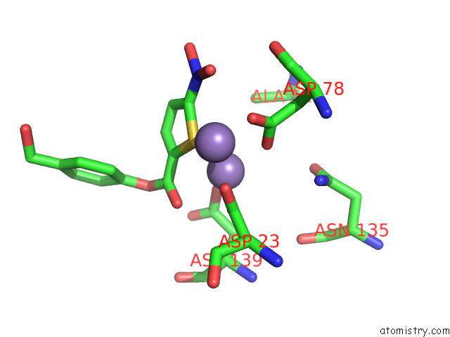
Mono view
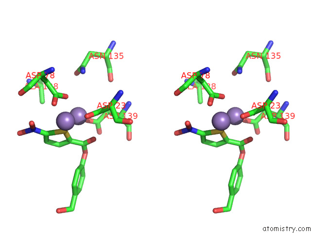
Stereo pair view
Mono view
Stereo pair view
A full contact list of Manganese with other atoms in the Mn binding
site number 2 of Crystal Structure of Engineered Hiv-1 Reverse Transcriptase Rnase H Domain Complexed with Nitrofuran Methoxy(Methoxycarbonyl)Phenyl Ester within 5.0Å range:
|
Reference:
H.Lu,
Y.Komukai,
K.Usami,
Y.Guo,
X.Qiao,
M.Nukaga,
T.Hoshino.
Computational and Crystallographic Analysis of Binding Structures of Inhibitory Compounds For Hiv-1 Rnase H Activity. J.Chem.Inf.Model. 2022.
ISSN: ESSN 1549-960X
PubMed: 36184946
DOI: 10.1021/ACS.JCIM.2C00537
Page generated: Sun Oct 6 11:05:47 2024
ISSN: ESSN 1549-960X
PubMed: 36184946
DOI: 10.1021/ACS.JCIM.2C00537
Last articles
Zn in 9MJ5Zn in 9HNW
Zn in 9G0L
Zn in 9FNE
Zn in 9DZN
Zn in 9E0I
Zn in 9D32
Zn in 9DAK
Zn in 8ZXC
Zn in 8ZUF