Manganese in PDB 7x9i: Crystal Structure of Mutt-8-Oxo-Dgtp Complex: Reaction For 12 Hr Using 5 Mm MN2+
Enzymatic activity of Crystal Structure of Mutt-8-Oxo-Dgtp Complex: Reaction For 12 Hr Using 5 Mm MN2+
All present enzymatic activity of Crystal Structure of Mutt-8-Oxo-Dgtp Complex: Reaction For 12 Hr Using 5 Mm MN2+:
3.6.1.55;
Protein crystallography data
The structure of Crystal Structure of Mutt-8-Oxo-Dgtp Complex: Reaction For 12 Hr Using 5 Mm MN2+, PDB code: 7x9i
was solved by
T.Nakamura,
Y.Yamagata,
with X-Ray Crystallography technique. A brief refinement statistics is given in the table below:
Resolution Low / High (Å)
|
32.14 /
1.90
|
Space group
|
P 21 21 21
|
Cell size a, b, c (Å), α, β, γ (°)
|
38.239,
56.004,
59.34,
90,
90,
90
|
R / Rfree (%)
|
14.4 /
19
|
Other elements in 7x9i:
The structure of Crystal Structure of Mutt-8-Oxo-Dgtp Complex: Reaction For 12 Hr Using 5 Mm MN2+ also contains other interesting chemical elements:
Manganese Binding Sites:
The binding sites of Manganese atom in the Crystal Structure of Mutt-8-Oxo-Dgtp Complex: Reaction For 12 Hr Using 5 Mm MN2+
(pdb code 7x9i). This binding sites where shown within
5.0 Angstroms radius around Manganese atom.
In total 3 binding sites of Manganese where determined in the
Crystal Structure of Mutt-8-Oxo-Dgtp Complex: Reaction For 12 Hr Using 5 Mm MN2+, PDB code: 7x9i:
Jump to Manganese binding site number:
1;
2;
3;
Manganese binding site 1 out
of 3 in 7x9i
Go back to
Manganese Binding Sites List in 7x9i
Manganese binding site 1 out
of 3 in the Crystal Structure of Mutt-8-Oxo-Dgtp Complex: Reaction For 12 Hr Using 5 Mm MN2+
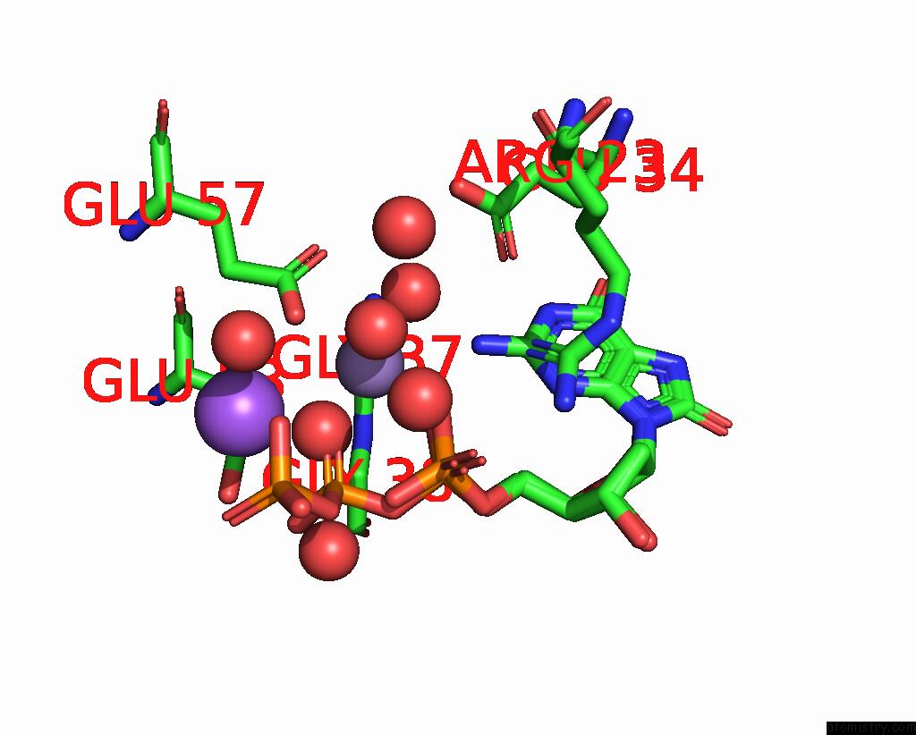
Mono view
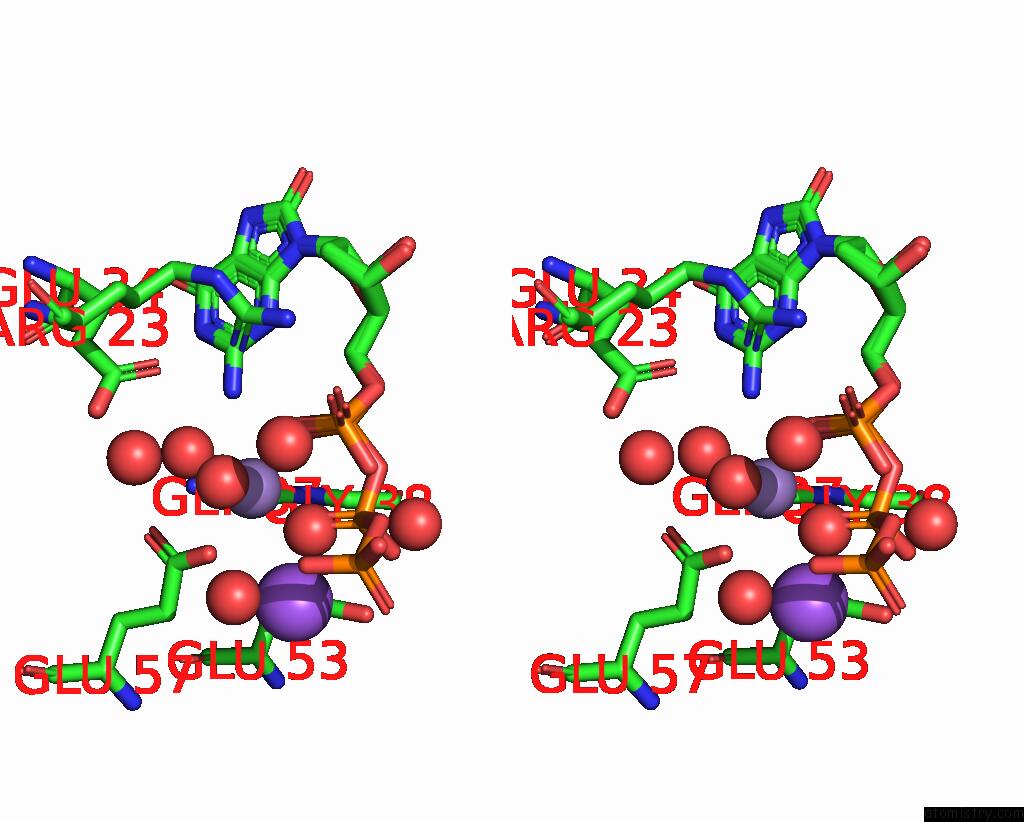
Stereo pair view
|
A full contact list of Manganese with other atoms in the Mn binding
site number 1 of Crystal Structure of Mutt-8-Oxo-Dgtp Complex: Reaction For 12 Hr Using 5 Mm MN2+ within 5.0Å range:
probe
|
atom
|
residue
|
distance (Å)
|
B
|
Occ
|
A:Mn204
b:12.5
occ:0.80
|
O2B
|
A:8DG202
|
2.1
|
15.9
|
0.3
|
O
|
A:GLY37
|
2.2
|
5.8
|
1.0
|
O
|
A:HOH408
|
2.2
|
16.4
|
0.7
|
O
|
A:HOH339
|
2.2
|
13.4
|
1.0
|
O1A
|
A:8DG202
|
2.2
|
10.0
|
0.3
|
OP2
|
A:8OG203
|
2.3
|
10.0
|
0.7
|
OE2
|
A:GLU57
|
2.3
|
14.0
|
1.0
|
O
|
A:HOH402
|
2.3
|
16.2
|
1.0
|
CD
|
A:GLU57
|
3.2
|
13.5
|
1.0
|
C
|
A:GLY37
|
3.4
|
11.4
|
1.0
|
PA
|
A:8DG202
|
3.4
|
12.5
|
0.3
|
P
|
A:8OG203
|
3.4
|
13.7
|
0.7
|
PB
|
A:8DG202
|
3.5
|
9.3
|
0.3
|
MN
|
A:MN206
|
3.5
|
30.4
|
0.3
|
OE1
|
A:GLU57
|
3.5
|
9.8
|
1.0
|
NA
|
A:NA207
|
3.5
|
30.9
|
0.7
|
OP1
|
A:8OG203
|
3.6
|
19.5
|
0.7
|
N2
|
A:8OG203
|
3.8
|
4.7
|
0.7
|
O3A
|
A:8DG202
|
3.8
|
10.4
|
0.3
|
N2
|
A:8DG202
|
3.9
|
4.8
|
0.3
|
O
|
A:HOH307
|
4.0
|
17.9
|
1.0
|
CA
|
A:GLY38
|
4.0
|
6.0
|
1.0
|
O2A
|
A:8DG202
|
4.1
|
11.1
|
0.3
|
N
|
A:GLY38
|
4.2
|
6.1
|
1.0
|
OP3
|
A:8OG203
|
4.3
|
11.1
|
0.7
|
N
|
A:GLY37
|
4.3
|
4.1
|
1.0
|
O
|
A:HOH347
|
4.3
|
19.6
|
1.0
|
O1G
|
A:8DG202
|
4.3
|
26.1
|
0.3
|
NH1
|
A:ARG23
|
4.4
|
7.1
|
1.0
|
O
|
A:HOH325
|
4.4
|
29.9
|
1.0
|
OE1
|
A:GLU53
|
4.4
|
9.0
|
1.0
|
CA
|
A:GLY37
|
4.5
|
4.9
|
1.0
|
O1B
|
A:8DG202
|
4.5
|
10.1
|
0.3
|
OE1
|
A:GLU34
|
4.5
|
8.0
|
1.0
|
O3B
|
A:8DG202
|
4.5
|
17.8
|
0.3
|
CG
|
A:GLU57
|
4.6
|
8.1
|
1.0
|
O5'
|
A:8OG203
|
4.6
|
13.2
|
0.7
|
O5'
|
A:8DG202
|
4.7
|
12.4
|
0.3
|
O
|
A:HOH431
|
4.9
|
12.0
|
0.7
|
C5'
|
A:8OG203
|
4.9
|
7.3
|
0.7
|
|
Manganese binding site 2 out
of 3 in 7x9i
Go back to
Manganese Binding Sites List in 7x9i
Manganese binding site 2 out
of 3 in the Crystal Structure of Mutt-8-Oxo-Dgtp Complex: Reaction For 12 Hr Using 5 Mm MN2+
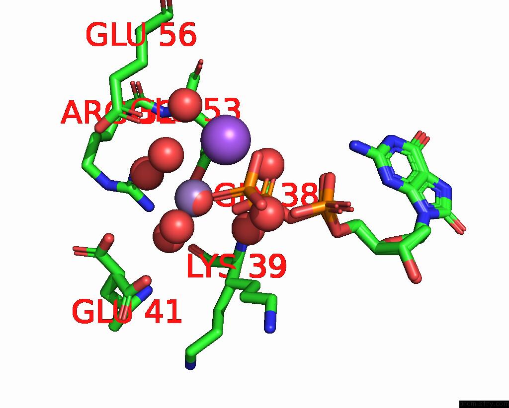
Mono view
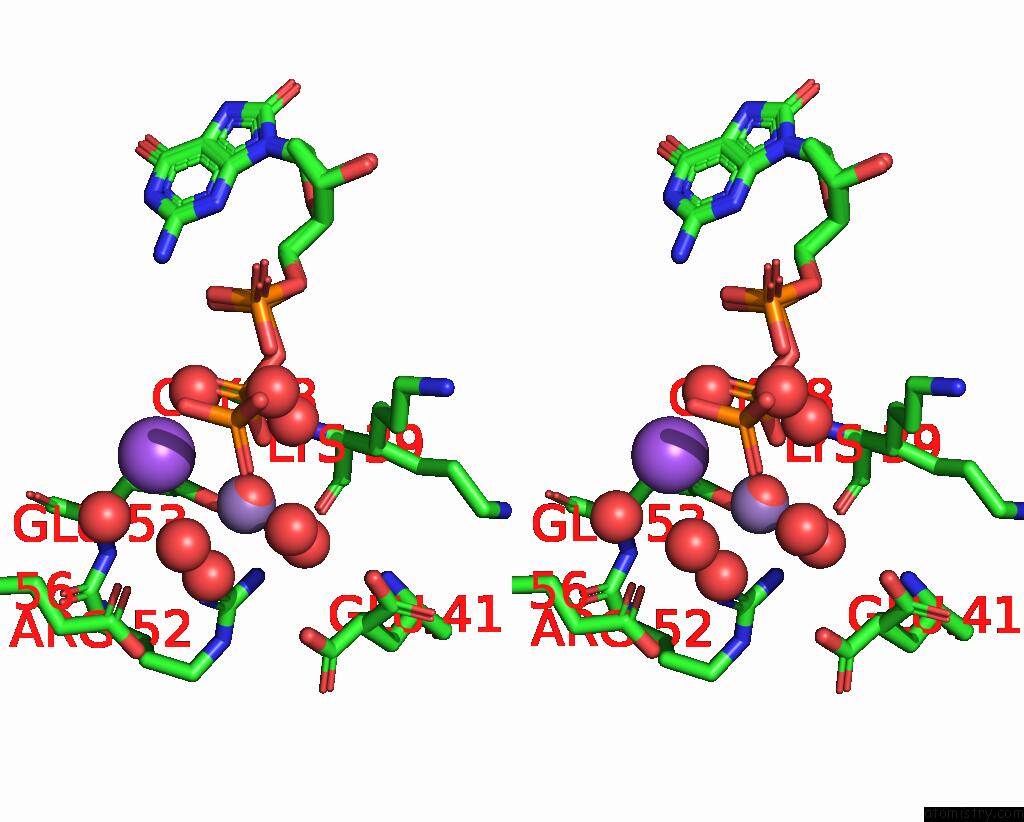
Stereo pair view
|
A full contact list of Manganese with other atoms in the Mn binding
site number 2 of Crystal Structure of Mutt-8-Oxo-Dgtp Complex: Reaction For 12 Hr Using 5 Mm MN2+ within 5.0Å range:
probe
|
atom
|
residue
|
distance (Å)
|
B
|
Occ
|
A:Mn205
b:21.0
occ:0.30
|
O
|
A:HOH355
|
0.2
|
21.2
|
0.7
|
O1B
|
A:8DG202
|
2.0
|
10.1
|
0.3
|
O
|
A:HOH323
|
2.4
|
20.3
|
1.0
|
O
|
A:HOH301
|
2.5
|
11.1
|
1.0
|
O
|
A:HOH302
|
2.6
|
20.5
|
0.3
|
OE2
|
A:GLU53
|
2.7
|
13.8
|
1.0
|
O
|
A:HOH431
|
2.9
|
12.0
|
0.7
|
NA
|
A:NA207
|
3.0
|
30.9
|
0.7
|
MN
|
A:MN206
|
3.2
|
30.4
|
0.3
|
O3G
|
A:8DG202
|
3.3
|
16.9
|
0.3
|
PB
|
A:8DG202
|
3.3
|
9.3
|
0.3
|
CD
|
A:GLU53
|
3.6
|
9.5
|
1.0
|
O
|
A:HOH408
|
3.7
|
16.4
|
0.7
|
OE1
|
A:GLU53
|
3.8
|
9.0
|
1.0
|
O
|
A:HOH351
|
3.9
|
36.4
|
1.0
|
O3B
|
A:8DG202
|
3.9
|
17.8
|
0.3
|
O2B
|
A:8DG202
|
4.0
|
15.9
|
0.3
|
NH1
|
A:ARG52
|
4.1
|
9.7
|
1.0
|
N
|
A:LYS39
|
4.2
|
6.9
|
0.4
|
N
|
A:LYS39
|
4.2
|
6.9
|
0.6
|
PG
|
A:8DG202
|
4.2
|
22.3
|
0.3
|
O
|
A:HOH311
|
4.4
|
15.8
|
1.0
|
OE1
|
A:GLU56
|
4.4
|
15.6
|
1.0
|
O3A
|
A:8DG202
|
4.5
|
10.4
|
0.3
|
OE2
|
A:GLU41
|
4.6
|
18.2
|
0.5
|
O
|
A:HOH447
|
4.6
|
35.6
|
0.7
|
OE2
|
A:GLU41
|
4.6
|
23.3
|
0.5
|
CA
|
A:GLY38
|
4.6
|
6.0
|
1.0
|
O
|
A:LYS39
|
4.7
|
7.0
|
0.6
|
O
|
A:LYS39
|
4.7
|
7.0
|
0.4
|
O1G
|
A:8DG202
|
4.7
|
26.1
|
0.3
|
OP1
|
A:8OG203
|
4.7
|
19.5
|
0.7
|
C
|
A:GLY38
|
4.8
|
8.5
|
1.0
|
CB
|
A:LYS39
|
4.9
|
11.7
|
0.4
|
NH2
|
A:ARG52
|
4.9
|
5.9
|
1.0
|
CB
|
A:LYS39
|
4.9
|
11.7
|
0.6
|
CZ
|
A:ARG52
|
5.0
|
8.8
|
1.0
|
|
Manganese binding site 3 out
of 3 in 7x9i
Go back to
Manganese Binding Sites List in 7x9i
Manganese binding site 3 out
of 3 in the Crystal Structure of Mutt-8-Oxo-Dgtp Complex: Reaction For 12 Hr Using 5 Mm MN2+
Mono view
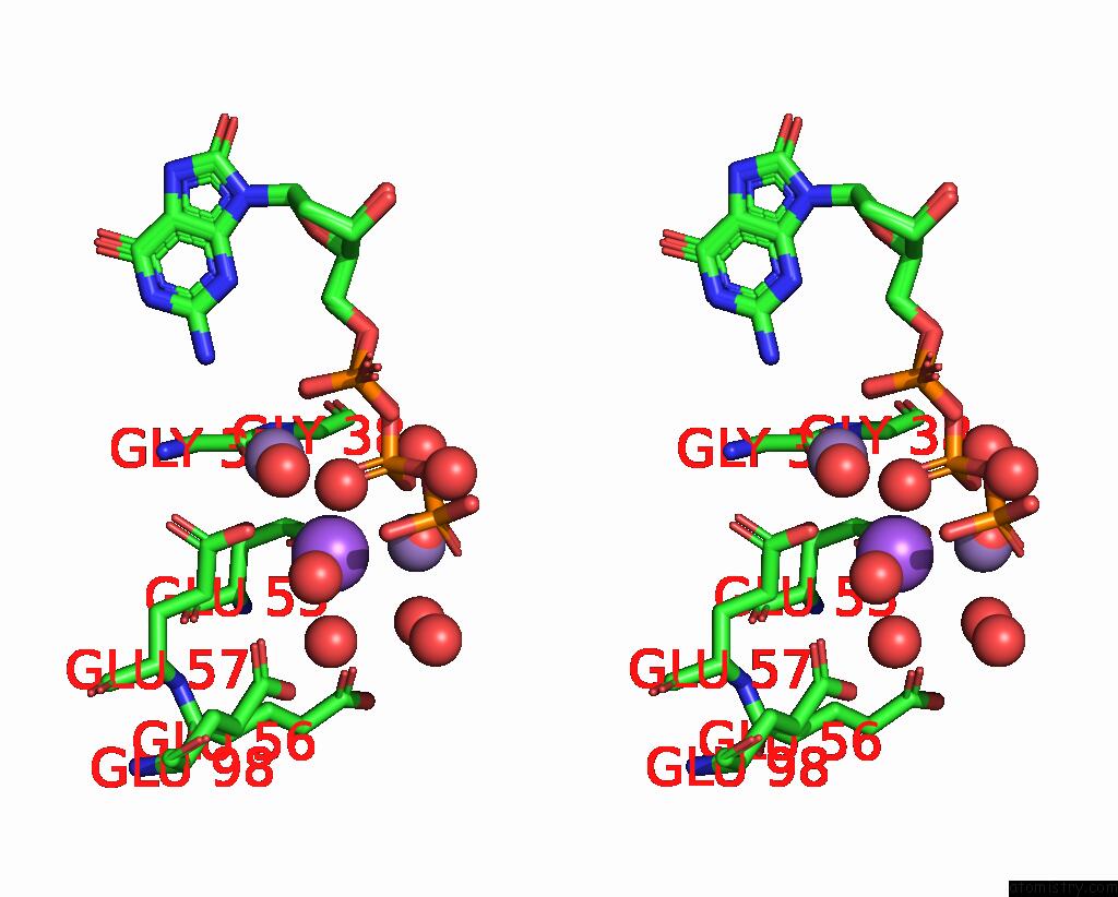
Stereo pair view
|
A full contact list of Manganese with other atoms in the Mn binding
site number 3 of Crystal Structure of Mutt-8-Oxo-Dgtp Complex: Reaction For 12 Hr Using 5 Mm MN2+ within 5.0Å range:
probe
|
atom
|
residue
|
distance (Å)
|
B
|
Occ
|
A:Mn206
b:30.4
occ:0.30
|
NA
|
A:NA207
|
0.2
|
30.9
|
0.7
|
O
|
A:HOH408
|
2.3
|
16.4
|
0.7
|
O
|
A:HOH311
|
2.5
|
15.8
|
1.0
|
OE1
|
A:GLU53
|
2.6
|
9.0
|
1.0
|
O2B
|
A:8DG202
|
2.6
|
15.9
|
0.3
|
OE2
|
A:GLU57
|
2.7
|
14.0
|
1.0
|
O
|
A:HOH307
|
2.8
|
17.9
|
1.0
|
O
|
A:HOH355
|
3.2
|
21.2
|
0.7
|
MN
|
A:MN205
|
3.2
|
21.0
|
0.3
|
PB
|
A:8DG202
|
3.4
|
9.3
|
0.3
|
O1B
|
A:8DG202
|
3.4
|
10.1
|
0.3
|
O1G
|
A:8DG202
|
3.5
|
26.1
|
0.3
|
MN
|
A:MN204
|
3.5
|
12.5
|
0.8
|
CD
|
A:GLU53
|
3.5
|
9.5
|
1.0
|
CD
|
A:GLU57
|
3.6
|
13.5
|
1.0
|
OE2
|
A:GLU53
|
3.7
|
13.8
|
1.0
|
CG
|
A:GLU57
|
3.7
|
8.1
|
1.0
|
O
|
A:GLY37
|
3.9
|
5.8
|
1.0
|
O
|
A:HOH323
|
4.0
|
20.3
|
1.0
|
O3G
|
A:8DG202
|
4.1
|
16.9
|
0.3
|
O
|
A:HOH351
|
4.1
|
36.4
|
1.0
|
OE1
|
A:GLU56
|
4.1
|
15.6
|
1.0
|
PG
|
A:8DG202
|
4.2
|
22.3
|
0.3
|
O3B
|
A:8DG202
|
4.3
|
17.8
|
0.3
|
O
|
A:HOH431
|
4.4
|
12.0
|
0.7
|
O
|
A:HOH402
|
4.4
|
16.2
|
1.0
|
OE2
|
A:GLU98
|
4.5
|
14.1
|
1.0
|
CA
|
A:GLY38
|
4.6
|
6.0
|
1.0
|
OE1
|
A:GLU98
|
4.7
|
16.0
|
1.0
|
OE1
|
A:GLU57
|
4.7
|
9.8
|
1.0
|
C
|
A:GLY37
|
4.7
|
11.4
|
1.0
|
OP1
|
A:8OG203
|
4.7
|
19.5
|
0.7
|
O3A
|
A:8DG202
|
4.8
|
10.4
|
0.3
|
O
|
A:HOH447
|
4.9
|
35.6
|
0.7
|
CG
|
A:GLU53
|
4.9
|
4.7
|
1.0
|
|
Reference:
T.Nakamura,
Y.Yamagata.
Visualization of Mutagenic Nucleotide Processing By Escherichia Coli Mutt, A Nudix Hydrolase. Proc.Natl.Acad.Sci.Usa V. 119 18119 2022.
ISSN: ESSN 1091-6490
PubMed: 35594391
DOI: 10.1073/PNAS.2203118119
Page generated: Sun Oct 6 11:04:54 2024
|