Manganese »
PDB 7uuh-7x9i »
7ww7 »
Manganese in PDB 7ww7: Crystal Structure of Mutt-8-Oxo-Dgtp Complex: Reaction For 1 Hr in 5 Mm MN2+
Enzymatic activity of Crystal Structure of Mutt-8-Oxo-Dgtp Complex: Reaction For 1 Hr in 5 Mm MN2+
All present enzymatic activity of Crystal Structure of Mutt-8-Oxo-Dgtp Complex: Reaction For 1 Hr in 5 Mm MN2+:
3.6.1.55;
3.6.1.55;
Protein crystallography data
The structure of Crystal Structure of Mutt-8-Oxo-Dgtp Complex: Reaction For 1 Hr in 5 Mm MN2+, PDB code: 7ww7
was solved by
T.Nakamura,
Y.Yamagata,
with X-Ray Crystallography technique. A brief refinement statistics is given in the table below:
| Resolution Low / High (Å) | 31.60 / 1.67 |
| Space group | P 21 21 21 |
| Cell size a, b, c (Å), α, β, γ (°) | 38.278, 55.968, 59.181, 90, 90, 90 |
| R / Rfree (%) | 15.9 / 19.8 |
Other elements in 7ww7:
The structure of Crystal Structure of Mutt-8-Oxo-Dgtp Complex: Reaction For 1 Hr in 5 Mm MN2+ also contains other interesting chemical elements:
| Sodium | (Na) | 1 atom |
Manganese Binding Sites:
The binding sites of Manganese atom in the Crystal Structure of Mutt-8-Oxo-Dgtp Complex: Reaction For 1 Hr in 5 Mm MN2+
(pdb code 7ww7). This binding sites where shown within
5.0 Angstroms radius around Manganese atom.
In total 3 binding sites of Manganese where determined in the Crystal Structure of Mutt-8-Oxo-Dgtp Complex: Reaction For 1 Hr in 5 Mm MN2+, PDB code: 7ww7:
Jump to Manganese binding site number: 1; 2; 3;
In total 3 binding sites of Manganese where determined in the Crystal Structure of Mutt-8-Oxo-Dgtp Complex: Reaction For 1 Hr in 5 Mm MN2+, PDB code: 7ww7:
Jump to Manganese binding site number: 1; 2; 3;
Manganese binding site 1 out of 3 in 7ww7
Go back to
Manganese binding site 1 out
of 3 in the Crystal Structure of Mutt-8-Oxo-Dgtp Complex: Reaction For 1 Hr in 5 Mm MN2+
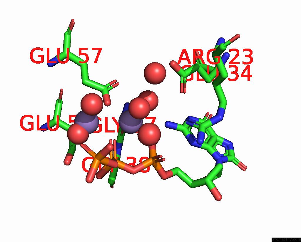
Mono view
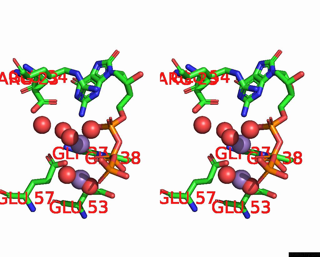
Stereo pair view
Mono view
Stereo pair view
A full contact list of Manganese with other atoms in the Mn binding
site number 1 of Crystal Structure of Mutt-8-Oxo-Dgtp Complex: Reaction For 1 Hr in 5 Mm MN2+ within 5.0Å range:
|
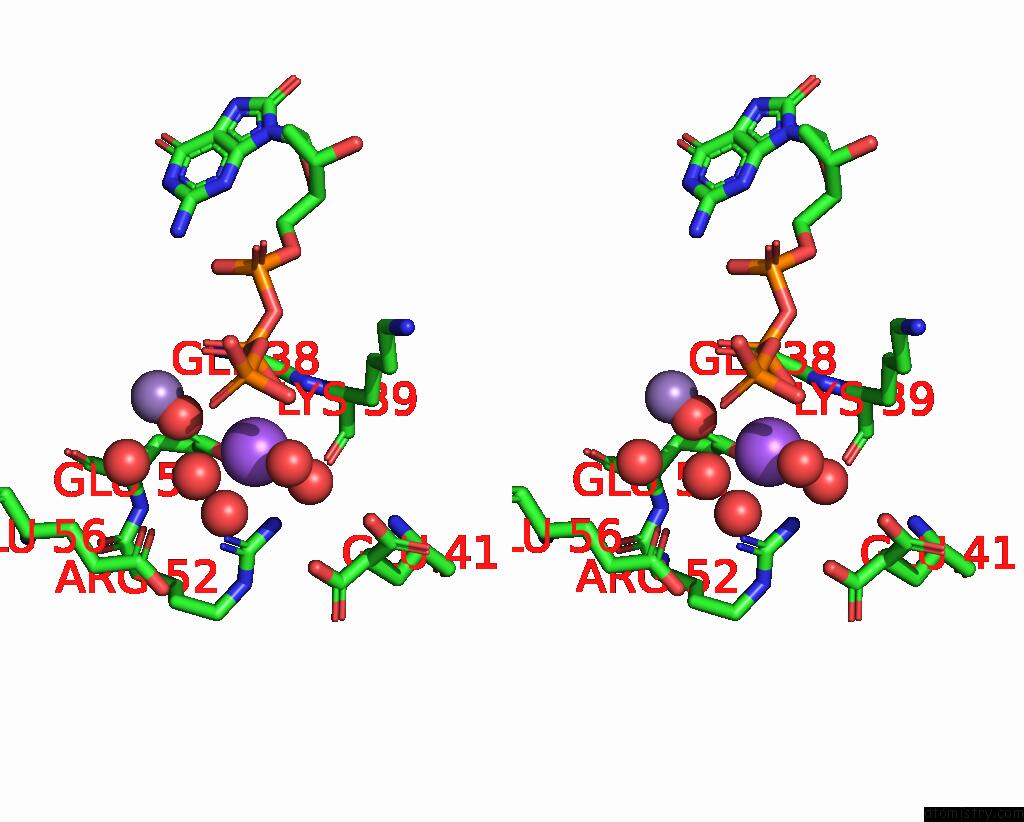
Manganese binding site 2 out of 3 in 7ww7
Go back to
Manganese binding site 2 out
of 3 in the Crystal Structure of Mutt-8-Oxo-Dgtp Complex: Reaction For 1 Hr in 5 Mm MN2+

Mono view
Stereo pair view
Mono view
Stereo pair view
A full contact list of Manganese with other atoms in the Mn binding
site number 2 of Crystal Structure of Mutt-8-Oxo-Dgtp Complex: Reaction For 1 Hr in 5 Mm MN2+ within 5.0Å range:
|
Manganese binding site 3 out of 3 in 7ww7
Go back to
Manganese binding site 3 out
of 3 in the Crystal Structure of Mutt-8-Oxo-Dgtp Complex: Reaction For 1 Hr in 5 Mm MN2+
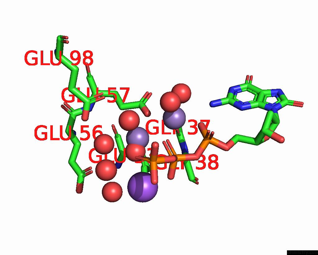
Mono view

Stereo pair view
Mono view
Stereo pair view
A full contact list of Manganese with other atoms in the Mn binding
site number 3 of Crystal Structure of Mutt-8-Oxo-Dgtp Complex: Reaction For 1 Hr in 5 Mm MN2+ within 5.0Å range:
|
Reference:
T.Nakamura,
Y.Yamagata.
Visualization of Mutagenic Nucleotide Processing By Escherichia Coli Mutt, A Nudix Hydrolase. Proc.Natl.Acad.Sci.Usa V. 119 18119 2022.
ISSN: ESSN 1091-6490
PubMed: 35594391
DOI: 10.1073/PNAS.2203118119
Page generated: Sun Oct 6 11:02:16 2024
ISSN: ESSN 1091-6490
PubMed: 35594391
DOI: 10.1073/PNAS.2203118119
Last articles
Zn in 9J0NZn in 9J0O
Zn in 9J0P
Zn in 9FJX
Zn in 9EKB
Zn in 9C0F
Zn in 9CAH
Zn in 9CH0
Zn in 9CH3
Zn in 9CH1