Manganese »
PDB 7uuh-7x9i »
7w5p »
Manganese in PDB 7w5p: Crystal Structure of the Dioxygenase Cctet From Coprinopsis Cinereain Bound to 12BP N6-Methyldeoxyadenine (6MA) Containing Duplex Dna
Protein crystallography data
The structure of Crystal Structure of the Dioxygenase Cctet From Coprinopsis Cinereain Bound to 12BP N6-Methyldeoxyadenine (6MA) Containing Duplex Dna, PDB code: 7w5p
was solved by
Y.J.Mu,
L.Zhang,
L.Zhang,
with X-Ray Crystallography technique. A brief refinement statistics is given in the table below:
| Resolution Low / High (Å) | 49.18 / 2.30 |
| Space group | P 21 21 21 |
| Cell size a, b, c (Å), α, β, γ (°) | 104.788, 105.198, 196.717, 90, 90, 90 |
| R / Rfree (%) | 22.8 / 25.7 |
Manganese Binding Sites:
The binding sites of Manganese atom in the Crystal Structure of the Dioxygenase Cctet From Coprinopsis Cinereain Bound to 12BP N6-Methyldeoxyadenine (6MA) Containing Duplex Dna
(pdb code 7w5p). This binding sites where shown within
5.0 Angstroms radius around Manganese atom.
In total 4 binding sites of Manganese where determined in the Crystal Structure of the Dioxygenase Cctet From Coprinopsis Cinereain Bound to 12BP N6-Methyldeoxyadenine (6MA) Containing Duplex Dna, PDB code: 7w5p:
Jump to Manganese binding site number: 1; 2; 3; 4;
In total 4 binding sites of Manganese where determined in the Crystal Structure of the Dioxygenase Cctet From Coprinopsis Cinereain Bound to 12BP N6-Methyldeoxyadenine (6MA) Containing Duplex Dna, PDB code: 7w5p:
Jump to Manganese binding site number: 1; 2; 3; 4;
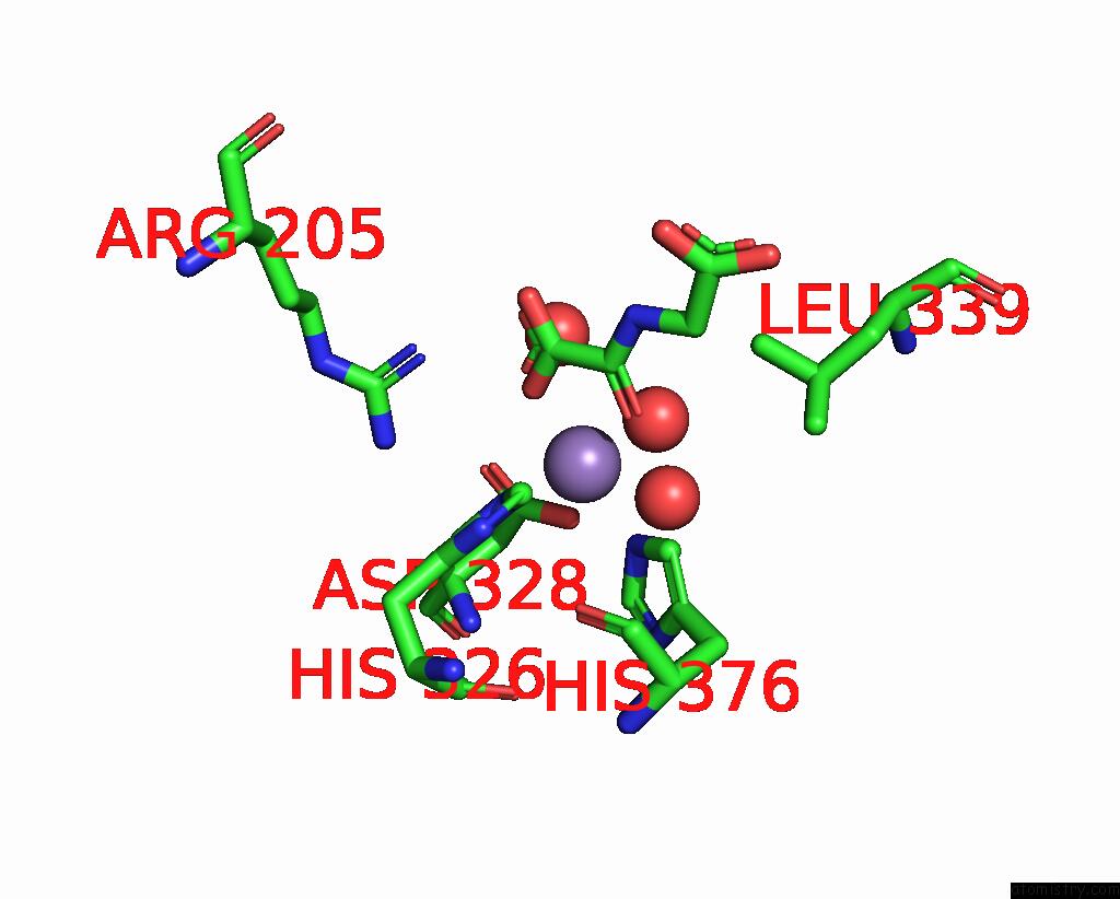
Manganese binding site 1 out of 4 in 7w5p
Go back to
Manganese binding site 1 out
of 4 in the Crystal Structure of the Dioxygenase Cctet From Coprinopsis Cinereain Bound to 12BP N6-Methyldeoxyadenine (6MA) Containing Duplex Dna
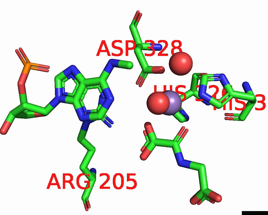
Mono view
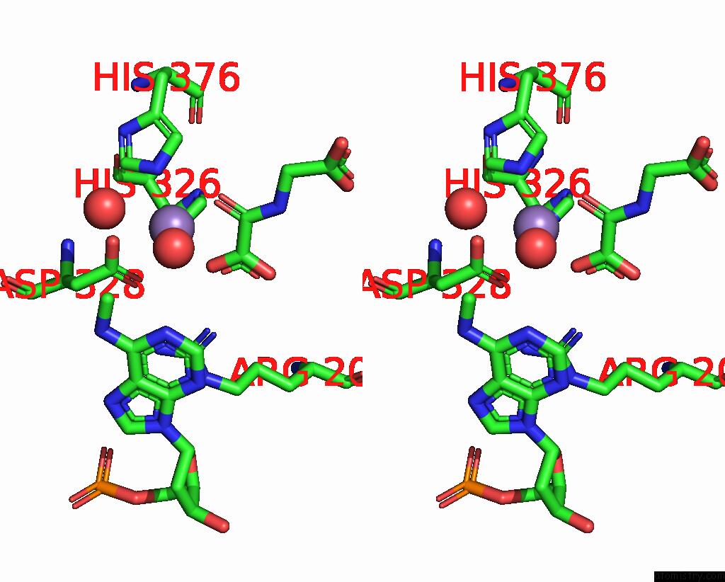
Stereo pair view
Mono view
Stereo pair view
A full contact list of Manganese with other atoms in the Mn binding
site number 1 of Crystal Structure of the Dioxygenase Cctet From Coprinopsis Cinereain Bound to 12BP N6-Methyldeoxyadenine (6MA) Containing Duplex Dna within 5.0Å range:
|
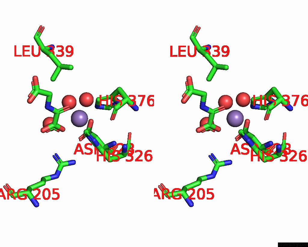
Manganese binding site 2 out of 4 in 7w5p
Go back to
Manganese binding site 2 out
of 4 in the Crystal Structure of the Dioxygenase Cctet From Coprinopsis Cinereain Bound to 12BP N6-Methyldeoxyadenine (6MA) Containing Duplex Dna
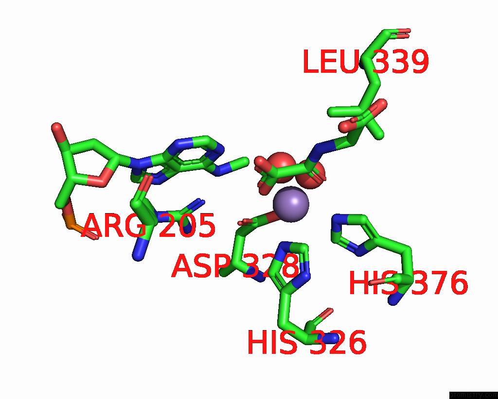
Mono view
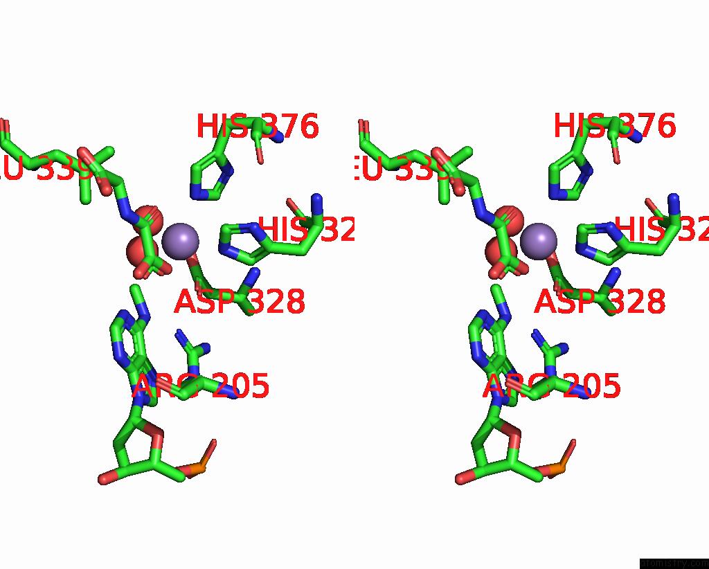
Stereo pair view
Mono view
Stereo pair view
A full contact list of Manganese with other atoms in the Mn binding
site number 2 of Crystal Structure of the Dioxygenase Cctet From Coprinopsis Cinereain Bound to 12BP N6-Methyldeoxyadenine (6MA) Containing Duplex Dna within 5.0Å range:
|
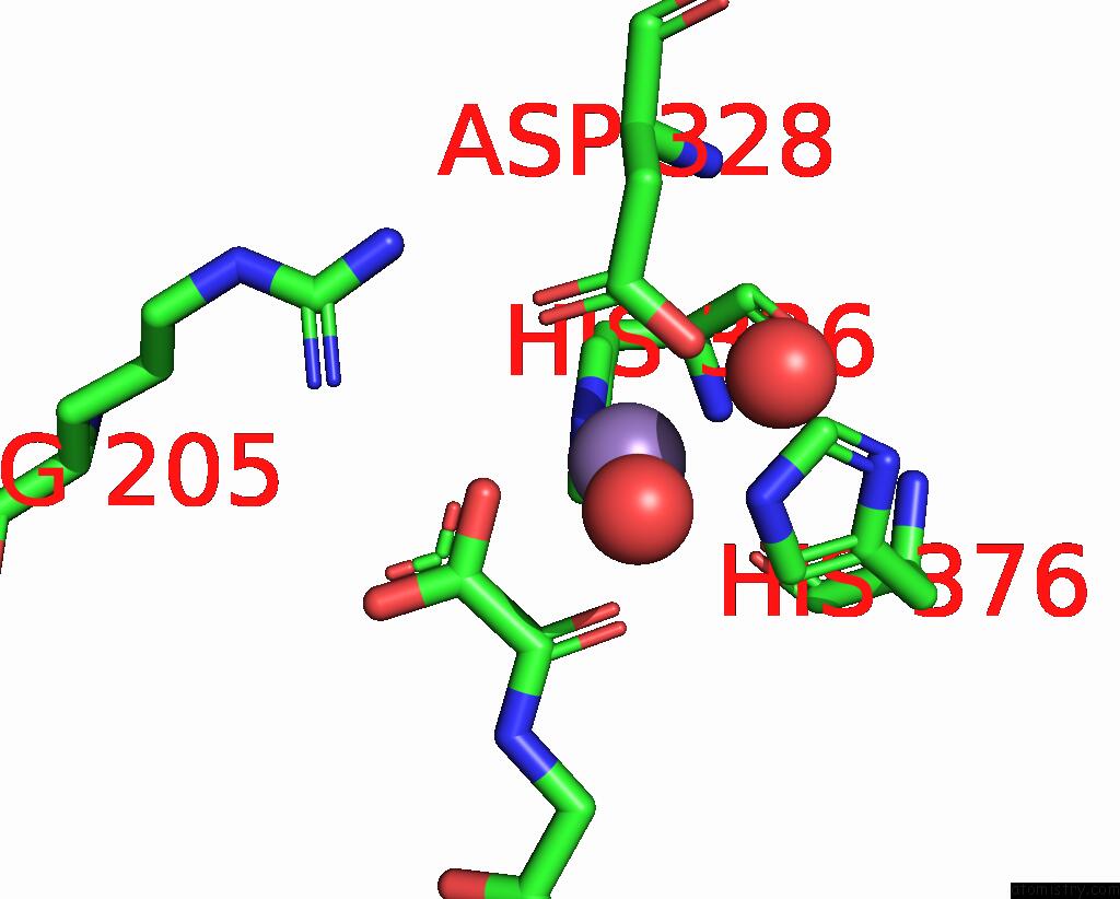
Manganese binding site 3 out of 4 in 7w5p
Go back to
Manganese binding site 3 out
of 4 in the Crystal Structure of the Dioxygenase Cctet From Coprinopsis Cinereain Bound to 12BP N6-Methyldeoxyadenine (6MA) Containing Duplex Dna
Mono view
Stereo pair view
Mono view
Stereo pair view
A full contact list of Manganese with other atoms in the Mn binding
site number 3 of Crystal Structure of the Dioxygenase Cctet From Coprinopsis Cinereain Bound to 12BP N6-Methyldeoxyadenine (6MA) Containing Duplex Dna within 5.0Å range:
|
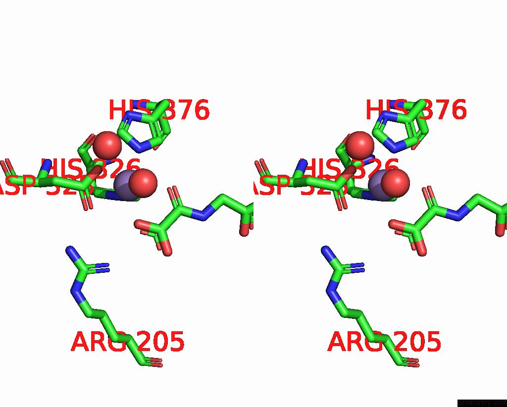
Manganese binding site 4 out of 4 in 7w5p
Go back to
Manganese binding site 4 out
of 4 in the Crystal Structure of the Dioxygenase Cctet From Coprinopsis Cinereain Bound to 12BP N6-Methyldeoxyadenine (6MA) Containing Duplex Dna
Mono view
Stereo pair view
Mono view
Stereo pair view
A full contact list of Manganese with other atoms in the Mn binding
site number 4 of Crystal Structure of the Dioxygenase Cctet From Coprinopsis Cinereain Bound to 12BP N6-Methyldeoxyadenine (6MA) Containing Duplex Dna within 5.0Å range:
|
Reference:
Y.Mu,
L.Zhang,
J.Hu,
J.Zhou,
H.W.Lin,
C.He,
H.Z.Chen,
L.Zhang.
A Fungal Dioxygenase Cctet Serves As A Eukaryotic 6MA Demethylase on Duplex Dna. Nat.Chem.Biol. V. 18 733 2022.
ISSN: ESSN 1552-4469
PubMed: 35654845
DOI: 10.1038/S41589-022-01041-3
Page generated: Sun Oct 6 10:59:13 2024
ISSN: ESSN 1552-4469
PubMed: 35654845
DOI: 10.1038/S41589-022-01041-3
Last articles
Zn in 9J0NZn in 9J0O
Zn in 9J0P
Zn in 9FJX
Zn in 9EKB
Zn in 9C0F
Zn in 9CAH
Zn in 9CH0
Zn in 9CH3
Zn in 9CH1