Manganese »
PDB 7u80-7utt »
7uq3 »
Manganese in PDB 7uq3: Jmjc Domain-Containing Protein 5 (JMJD5) in Complex with Mn and (S)-2- (1-Hydroxy-2,5-Dioxopyrrolidin-3-Yl)Acetic Acid
Enzymatic activity of Jmjc Domain-Containing Protein 5 (JMJD5) in Complex with Mn and (S)-2- (1-Hydroxy-2,5-Dioxopyrrolidin-3-Yl)Acetic Acid
All present enzymatic activity of Jmjc Domain-Containing Protein 5 (JMJD5) in Complex with Mn and (S)-2- (1-Hydroxy-2,5-Dioxopyrrolidin-3-Yl)Acetic Acid:
1.14.11.73;
1.14.11.73;
Protein crystallography data
The structure of Jmjc Domain-Containing Protein 5 (JMJD5) in Complex with Mn and (S)-2- (1-Hydroxy-2,5-Dioxopyrrolidin-3-Yl)Acetic Acid, PDB code: 7uq3
was solved by
R.Chowdhury,
M.S.Islam,
C.J.Schofield,
with X-Ray Crystallography technique. A brief refinement statistics is given in the table below:
| Resolution Low / High (Å) | 49.85 / 1.49 |
| Space group | P 21 21 21 |
| Cell size a, b, c (Å), α, β, γ (°) | 49.479, 64.69, 78.23, 90, 90, 90 |
| R / Rfree (%) | 13.4 / 13.5 |
Manganese Binding Sites:
The binding sites of Manganese atom in the Jmjc Domain-Containing Protein 5 (JMJD5) in Complex with Mn and (S)-2- (1-Hydroxy-2,5-Dioxopyrrolidin-3-Yl)Acetic Acid
(pdb code 7uq3). This binding sites where shown within
5.0 Angstroms radius around Manganese atom.
In total only one binding site of Manganese was determined in the Jmjc Domain-Containing Protein 5 (JMJD5) in Complex with Mn and (S)-2- (1-Hydroxy-2,5-Dioxopyrrolidin-3-Yl)Acetic Acid, PDB code: 7uq3:
In total only one binding site of Manganese was determined in the Jmjc Domain-Containing Protein 5 (JMJD5) in Complex with Mn and (S)-2- (1-Hydroxy-2,5-Dioxopyrrolidin-3-Yl)Acetic Acid, PDB code: 7uq3:
Manganese binding site 1 out of 1 in 7uq3
Go back to
Manganese binding site 1 out
of 1 in the Jmjc Domain-Containing Protein 5 (JMJD5) in Complex with Mn and (S)-2- (1-Hydroxy-2,5-Dioxopyrrolidin-3-Yl)Acetic Acid
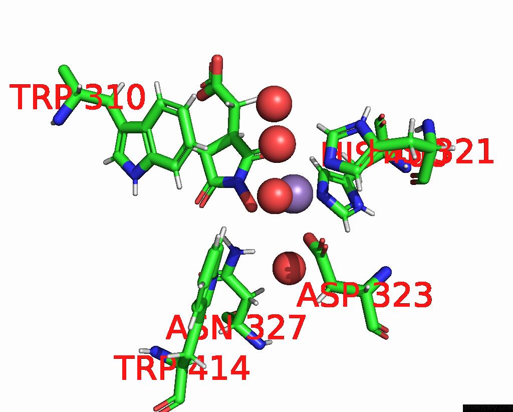
Mono view
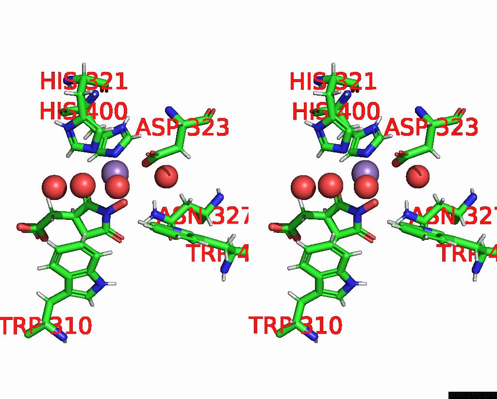
Stereo pair view
Mono view
Stereo pair view
A full contact list of Manganese with other atoms in the Mn binding
site number 1 of Jmjc Domain-Containing Protein 5 (JMJD5) in Complex with Mn and (S)-2- (1-Hydroxy-2,5-Dioxopyrrolidin-3-Yl)Acetic Acid within 5.0Å range:
|
Reference:
M.S.Islam,
M.Markoulides,
R.Chowdhury,
C.J.Schofield.
Structural Analysis of the 2-Oxoglutarate Binding Site of the Circadian Rhythm Linked Oxygenase JMJD5. Sci Rep V. 12 20680 2022.
ISSN: ESSN 2045-2322
PubMed: 36450832
DOI: 10.1038/S41598-022-24154-0
Page generated: Sun Oct 6 10:56:40 2024
ISSN: ESSN 2045-2322
PubMed: 36450832
DOI: 10.1038/S41598-022-24154-0
Last articles
Zn in 9J0NZn in 9J0O
Zn in 9J0P
Zn in 9FJX
Zn in 9EKB
Zn in 9C0F
Zn in 9CAH
Zn in 9CH0
Zn in 9CH3
Zn in 9CH1