Manganese »
PDB 7u80-7utt »
7ug9 »
Manganese in PDB 7ug9: Crystal Structure of Rnase Am Php Domain
Protein crystallography data
The structure of Crystal Structure of Rnase Am Php Domain, PDB code: 7ug9
was solved by
S.K.Doamekpor,
L.Tong,
with X-Ray Crystallography technique. A brief refinement statistics is given in the table below:
| Resolution Low / High (Å) | 37.19 / 1.69 |
| Space group | P 1 21 1 |
| Cell size a, b, c (Å), α, β, γ (°) | 40.289, 74.389, 40.079, 90, 118.98, 90 |
| R / Rfree (%) | 18.2 / 21.5 |
Manganese Binding Sites:
The binding sites of Manganese atom in the Crystal Structure of Rnase Am Php Domain
(pdb code 7ug9). This binding sites where shown within
5.0 Angstroms radius around Manganese atom.
In total 3 binding sites of Manganese where determined in the Crystal Structure of Rnase Am Php Domain, PDB code: 7ug9:
Jump to Manganese binding site number: 1; 2; 3;
In total 3 binding sites of Manganese where determined in the Crystal Structure of Rnase Am Php Domain, PDB code: 7ug9:
Jump to Manganese binding site number: 1; 2; 3;
Manganese binding site 1 out of 3 in 7ug9
Go back to
Manganese binding site 1 out
of 3 in the Crystal Structure of Rnase Am Php Domain

Mono view
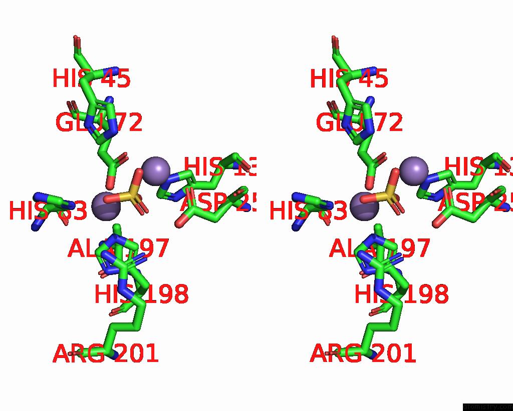
Stereo pair view
Mono view
Stereo pair view
A full contact list of Manganese with other atoms in the Mn binding
site number 1 of Crystal Structure of Rnase Am Php Domain within 5.0Å range:
|
Manganese binding site 2 out of 3 in 7ug9
Go back to
Manganese binding site 2 out
of 3 in the Crystal Structure of Rnase Am Php Domain

Mono view
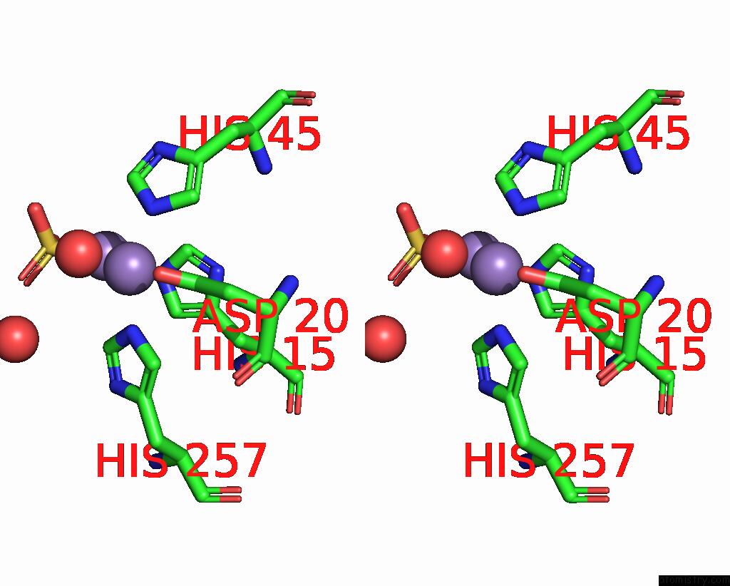
Stereo pair view
Mono view
Stereo pair view
A full contact list of Manganese with other atoms in the Mn binding
site number 2 of Crystal Structure of Rnase Am Php Domain within 5.0Å range:
|
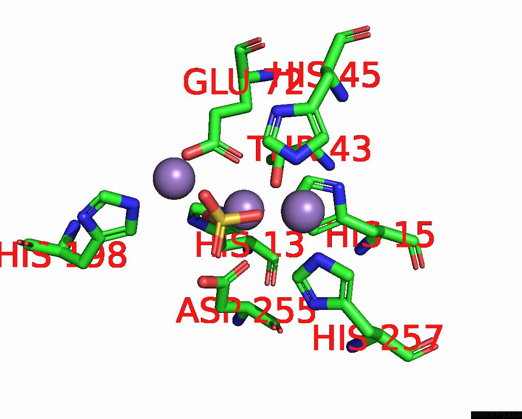
Manganese binding site 3 out of 3 in 7ug9
Go back to
Manganese binding site 3 out
of 3 in the Crystal Structure of Rnase Am Php Domain
Mono view

Stereo pair view
Mono view
Stereo pair view
A full contact list of Manganese with other atoms in the Mn binding
site number 3 of Crystal Structure of Rnase Am Php Domain within 5.0Å range:
|
Reference:
S.Sharma,
J.Yang,
S.K.Doamekpor,
E.Grudizen-Nogalska,
L.Tong,
M.Kiledjian.
Identification of A Novel Defadding Activity in Human, Yeast and Bacterial 5' to 3' Exoribonucleases. Nucleic Acids Res. V. 50 8807 2022.
ISSN: ESSN 1362-4962
PubMed: 35904778
DOI: 10.1093/NAR/GKAC617
Page generated: Sun Oct 6 10:50:49 2024
ISSN: ESSN 1362-4962
PubMed: 35904778
DOI: 10.1093/NAR/GKAC617
Last articles
Fe in 2W3HFe in 2W3G
Fe in 2W3F
Fe in 2W3E
Fe in 2W31
Fe in 2W3D
Fe in 2VR0
Fe in 2VXI
Fe in 2W23
Fe in 2W16